Posterior Ankle Impingement Syndrome
Posterior Ankle Impingement Syndrome
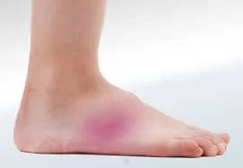
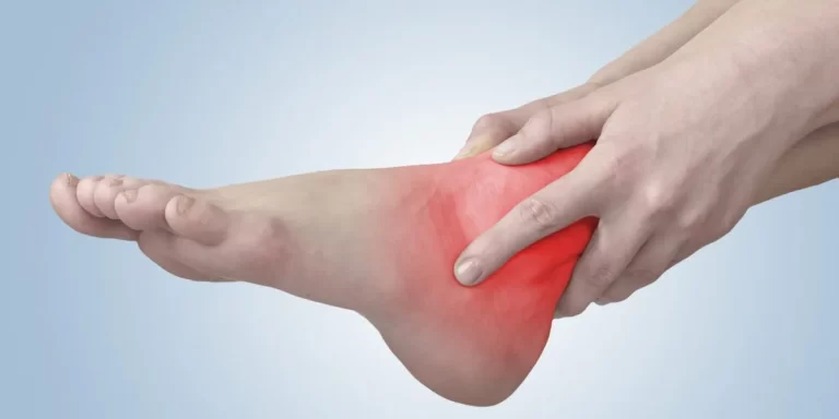
Posterior Ankle Impingement Syndrome (PAIS) is a condition characterized by pain at the back of the ankle, typically caused by compression of soft tissues or bony structures during activities that involve repetitive or forceful ankle plantarflexion, such as dancing, running, or kicking sports.
It often occurs due to anatomical variations like an os trigonum (an accessory bone) or thickened soft tissues. PAIS is common among athletes and can lead to chronic discomfort and reduced performance if not managed properly.
What is Ankle Impingement?
The term “ankle impingement” describes a condition where the ankle joint is painful and has limited range of motion. It happens when specific movements cause the soft tissues or bone structures inside the ankle to compress or pinch. This may result in joint pain and inflammation.
These two types of ankle impingement are referred to as anterior and posterior, respectively. Ankle structures in the front are compressed by anterior impingement, often known as “footballer’s ankle” or “athlete’s ankle.” This condition is usually caused by repeated ankle dorsiflexion or forced plantar flexion. The back of the ankle is compressed in posterior impingement, on the other hand, which is frequently caused by excessive plantar flexion.
Ankle impingement may be caused by abnormalities in the ankle joint, bone spurs, excessive straining on the joint, or past ankle injuries. Pain, stiffness, edema, and a reduced range of motion are possible symptoms.
Anterior & Posterior Ankle Impingement Syndrome
The symptoms of anterior and posterior ankle impingement syndromes include reduced range of motion and pain in the ankle joint. These disorders are caused by the tissues in the front or back of the ankle being compressed or pinched, respectively.
Anterior ankle impingement syndrome: This condition typically arises from recurrent compression of the soft tissues or bone structures near the front of the ankle. Activities involving forceful dorsiflexion (raising the foot) or recurrent ankle sprains may be the cause of this. This may eventually cause pain, inflammation, and a reduced ankle range of motion.
Posterior ankle impingement syndrome: Compression of the bone or soft tissues at the back of the ankle is known as posterior ankle impingement syndrome, and it is usually caused by repetitive plantar flexion, or lowering the foot. Activities like ballet and running, which require a lot of powerful push-off movements, can demonstrate this. The compression may cause pain and limited ankle motion, especially when doing tasks that require forced plantar flexion.
Conservative treatments include rest, ice, physical therapy exercises, and anti-inflammatory drugs to lessen discomfort and inflammation may be the first line of treatment for anterior and posterior ankle impingement syndromes. Additional therapies might be explored if symptoms worsen or continue. Corticosteroid injections, orthotics, activity adjustments, and, in extreme situations, surgery to remove bone deformities or repair damaged tissues, are a few examples of these.
What is the cause of posterior impingement syndrome?
Posterior impingement syndrome is caused by compression of the structures between the tibia, the bottom of the shin bone, and the talus, the bone that makes up the lower portion of the ankle joint. When the foot is plantarflexed, as happens when you stand on your tiptoes, the structures at the back of the ankle may compress between the back of the talus and the back of the tibia. Within these structures, this compression may result in edema and inflammation.
Anytime you place your foot in a toe-pointed position, these swelling structures may compress. Ballet and kicking sports are two examples of repetitive foot-pointing activities or sports that might result in posterior impingement syndrome. A bony abnormality in your ankle or an expanded bony prominence on the back of your foot bone, known as the “os trigonum,” can also result in posterior impingement.
Causes and Main Symptoms of the Posteromedial Ligament
The deltoid ligament, sometimes referred to as the posteromedial ligament, is a strong band of connective tissue that runs down the inside of the ankle. It keeps the foot from rolling too much inward or everting, and it stabilizes the ankle joint. Damage to the posteromedial ligament can result in a condition called deltoid ligament injury or posteromedial ligament injury.
Posteromedial ligament damage is primarily caused by:
Ankle sprains: Usually caused by activities involving sudden direction changes or uneven surfaces, these injuries can cause the ligament to strain or tear when the ankle is forcefully twisted or rolled inward.
Trauma or impact: The posteromedial ligament may sustain damage from direct trauma or a violent blow to the inside side of the ankle.
The following are some of the main signs of a posteromedial ligament injury:
- Tenderness and discomfort: The inner part of the ankle usually hurts, and this pain gets greater when you bear weight or move your foot inward.
- Swelling and bruising: Because of bleeding or inflammation, the damaged region may swell and exhibit bruises.
- Instability: Activities that put stress on the ligament can lead to ankle instability or a giving-way sensation.
- Restricted range of motion: Foot movements involving inversion or inward rotation may be uncomfortable or limited.
Mechanism of Injury
Acute traumatic plantar hyperflexion incidents, including ankle sprains, may be made worse by posterior ankle impingement. The female ballet dancer’s recurrent low-grade injuries associated with plantar hyperflexion may possibly be a contributing factor. Because in part to additional injuries that may be acquired during an acute traumatic incident, posterior impingement from overuse has a better prognosis; hence, it is crucial to distinguish between the two.
The occurrence of posterior impingement syndrome is significantly influenced by the structure of the ankle’s posterior portion. The os trigonum, an osteophyte from the posterior distal tibia, a large posterior process of the calcaneus, a downward-sloping posterior lip of the tibia, or a protracted posterolateral tubercle of the talus (Stieda’s process) are some of the more common osseous reasons.
Posttraumatic scar tissue, posttraumatic calcifications of the posterior joint capsule, loose bodies in the posterior portion of the ankle joint, and a thicker posterior joint capsule are some examples of soft tissue-related causes of posterior impingement. Compression of any of these structures can occur during plantar hyperflexion.
The posterior talus process may affect the posterior tibial plateau or the posterior calcaneal process, or the soft tissues between the two opposing osseous structures may compress, causing pain. All of these disorders cause painful limitations in ankle range of motion due to physical impingement of soft tissue or osseous structures.
The most common cause of soft tissue impingement is fibrosis and scarring caused by ligamentous, capsular, or synovial damage. The posterior capsule and the posterior talofibular, intermalleolar, and tibiofibular ligaments are among the capsular soft tissues involved in the context of posterior ankle impingement.
Clinical manifestation of the disease is believed to typically occur when a substantial soft-tissue component develops. The soft-tissue component may be more specific, affecting the talofibular ligament or posterior intermalleolar ligament, or it may be composed of synovial thickening across the posterior capsule. Stenosing tenosynovitis can arise from posterior impingement injuries to the flexor hallucis longus tendon, which runs in the groove between the lateral and medial processes of the talus.
Symptoms of posterior impingement syndrome
Posterior impingement syndrome causes pain in the back of the ankle when you point your foot. Other signs and symptoms could be:
- decreased mobility
- Weakness
- edema and inflammation
- balance issues
What is the diagnosis for posterior ankle impingement?
Clinical Presentation
Patients with posterior impingement experience persistent deep posterior ankle pain that is exacerbated by push-off pressures or forced plantar flexion, which can happen when jumping, jogging downhill, or ballet dancing. Soft tissue or bony structures may be compressed in these areas between the posterior tibia and the posterolateral process of the talus or between the posterior aspect of the distal tibia and the calcaneus. Forcible dorsiflexion may result in pain for certain persons. The posterior joint capsule and posterior talofibular ligament, which both connect to the posterolateral talar process, are subjected to traction in this dorsiflexed position.
The posterolateral talar process, which runs along the posterolateral portion of the ankle between the Achilles and peroneal tendons, feels painful when palpated during a physical examination. Because the posterolateral talar process is wedged between the posterior tibia and calcaneus, passive forced plantar flexion causes pain and often an uncomfortable feeling. If pain subsides after an anesthetic injection into the tibiotalar joint’s posterolateral capsule, the diagnosis is confirmed.
A thorough examination by a medical expert, usually an orthopaedic specialist or podiatrist, is necessary to diagnose posterior ankle impingement. The following can be part of the diagnostic procedure:
Medical history: To comprehend the background of your illness, the doctor will talk about your symptoms, past ankle injuries, and any relevant medical disorders.
Physical examination: Your ankle will be thoroughly examined by the medical professional to determine its range of motion, discomfort, edema, and tenderness. Additionally, they could assess the alignment of your ankle and foot and carry out particular movements to replicate symptoms.
Imaging tests: To check for any bony abnormalities that might be causing the impingement, such as bone spurs or joint degeneration, X-rays may be done. Soft tissues, such as ligaments, tendons, and joint structures, can be examined using magnetic resonance imaging (MRI) or ultrasound to check for inflammation or other structural abnormalities.
Diagnostic injection: To determine the cause of pain, a diagnostic injection may be used in certain situations. If the pain is related to impingement, injecting a corticosteroid or anesthetic into the area of pain can reduce inflammation or temporarily numb the area.
The medical practitioner can definitively diagnose posterior ankle impingement based on the results of the evaluation, physical examination, and imaging studies. In order to reduce symptoms and enhance ankle function, this diagnosis will direct the course of treatment and identify the best course of action.
For a precise diagnosis and a customized treatment plan suited to your particular disease, it is crucial to speak with a licensed healthcare provider.
Differential Diagnosis
The clinical condition known as posteromedial ankle impingement syndrome is comparable to posterior ankle impingement. After suffering a severe ankle inversion injury that crushed the deep posterior fibers of the deltoid ligament complex between the medial talus and the medial malleolus, the patient in this circumstance has chronic posteromedial ankle pain.
Immature scarring of the posterior talotibial ligament, which extends slightly into the posteromedial gutter, occurs after the initial edema. Posttraumatic synovitis is also present, thickening and shifting the posteromedial ankle capsule.
This results in fibrosis and scar tissue development along the posteromedial joint line, which gets trapped between the medial malleolus and the posterior talus. Although posteromedial ankle impingement can happen by itself, anterolateral ankle soreness and instability sensations are the most common co-occurring conditions. Following lateral ankle reconstruction, posteromedial ankle impingement may cause persistent discomfort if the lesion is not identified and resolved.
The posterior talotibial ligament usually loses its characteristic striated appearance on axial MR imaging, and synovitis and scar response expand posteriorly into the medial gutter. Moreover, the typical gap between the flexor digitorum longus and flexor hallucis longus tendon levels is eliminated, and the posteromedial ankle capsule thickens.
Many other conditions, such as talus and calcaneal fractures, Achilles tendinopathy, isolated flexor hallucis longus tendinopathy, retrocalcaneal bursitis, Haglund’s deformity, posterior tibial osteochondral injuries, tarsal coalition, and tarsal tunnel syndrome, may seem like posterior impingement.
Treatment For Posterior Ankle Impingement Syndrome
The degree of the impingement, a person’s capacity for healing, and the treatment strategy selected can all affect how long it takes to recover from an ankle impingement. Ankle impingement typically requires a few weeks to many months to recover from.
Conservative therapy is helpful for people who experience ankle impingement. Anti-inflammatory drugs, physical therapy exercises, and rest may be enough. In these situations, people frequently feel better and resume their normal activities in a few weeks.
Surgery may be necessary for severe diseases or in situations when conservative therapy is not working. Although recovery times after ankle impingement surgery can vary, they usually require a longer period of therapy. Physical therapy and progressive rehabilitation activities may be required to regain ankle strength, flexibility, and function, as surgical incisions can take weeks to heal. Recovery after surgery could take many months.
It is crucial to remember that every person’s healing process is unique and can be impacted by different things. A quicker and more successful recovery can be achieved by adhering to rehabilitation protocols, accepting lifestyle changes advised by your healthcare team, and following professional advice.
It is best to speak with a healthcare provider who can assess your unique situation and offer specific guidance depending on your circumstances in order to obtain a more precise estimate of recovery time for ankle impingement.
Physiotherapy treatment for posterior impingement syndrome.
Physical therapy is an important part of treating posterior impingement syndrome. First, your physiotherapist can determine the nature of your issue and how serious it is. Referrals for X-rays may be necessary in certain circumstances. Your physiotherapist will determine an appropriate treatment plan after diagnosing you. The course of treatment may involve the following:
- Electrotherapy
- Orthotics
- Hydrotherapy
- Manipulation / Mobilisation
Does posterior impingement syndrome have possible long-term effects?
When posterior impingement syndrome is correctly diagnosed and treated, there are no long-term consequences. However, in certain cases, surgery may be necessary to eliminate the impingement’s cause.
FAQs
What is the posterior ankle impingement injury mechanism?
Compression of the tissues posterior to the tibiotalar and talocalcaneal articulations during terminal plantar flexion causes posterior ankle impingement. Mechanical restriction from osteophytes and/or trapping of different soft tissue structures from inflammation, scarring, or hypermobility are the two main causes of pain.
How long does it take to recover from a posterior ankle impingement?
Once the impingement has become chronic, it is usually possible to return to sports and relieve discomfort by removing the inflammatory issue and abnormal bone. It normally takes three to six months to fully recover.
When you have a posterior ankle impingement, how do you exercise?
The main goals of exercises for posterior ankle impingement are to increase ankle range of motion gradually, strengthen supporting muscles, and increase flexibility. Heel rises, toe flexions, and calf stretches are typical exercises. Resistance bands are used in some activities to increase plantarflexion and stretch the ankle joint. To ensure correct technique and avoid future injury, it is essential to carry out these exercises under the supervision of a medical professional or physical therapist.
How to fix posterior ankle impingement?
Patients with posterior ankle impingement typically don’t need surgery. To reduce swelling and pain, you should use a compression bandage, elevate the ankle above your heart whenever possible, apply an ice pack frequently, and get lots of rest.
How to relieve foot pain in 30 seconds?
To ease foot pain, try stretching your feet for 30 seconds. Gently draw your toes towards your shin while placing one foot on top of the other. Maintain the stretch for 15–20 seconds. The plantar fascia is stretched, and this might offer immediate relief.
References:
- Ankle, C. F. &. (2023, June 28). Unlocking ankle freedom: posterior ankle impingement explained. Certified Foot & Ankle Specialists. https://certifiedfoot.com/unlocking-ankle-freedom-posterior-ankle-impingement-explained/
- Sharma, R., & Weerakkody, Y. (2010). Posterior ankle impingement syndrome. Radiopaedia.org. https://doi.org/10.53347/rid-10754
- Posterior impingement syndrome – ankle – conditions – musculoskeletal – What we treat – physio.co.uk. (n.d.). https://www.physio.co.uk/what-we-treat/musculoskeletal/conditions/ankle/posterior-impingement-syndrome.php



One Comment