Talus Bone
Introduction
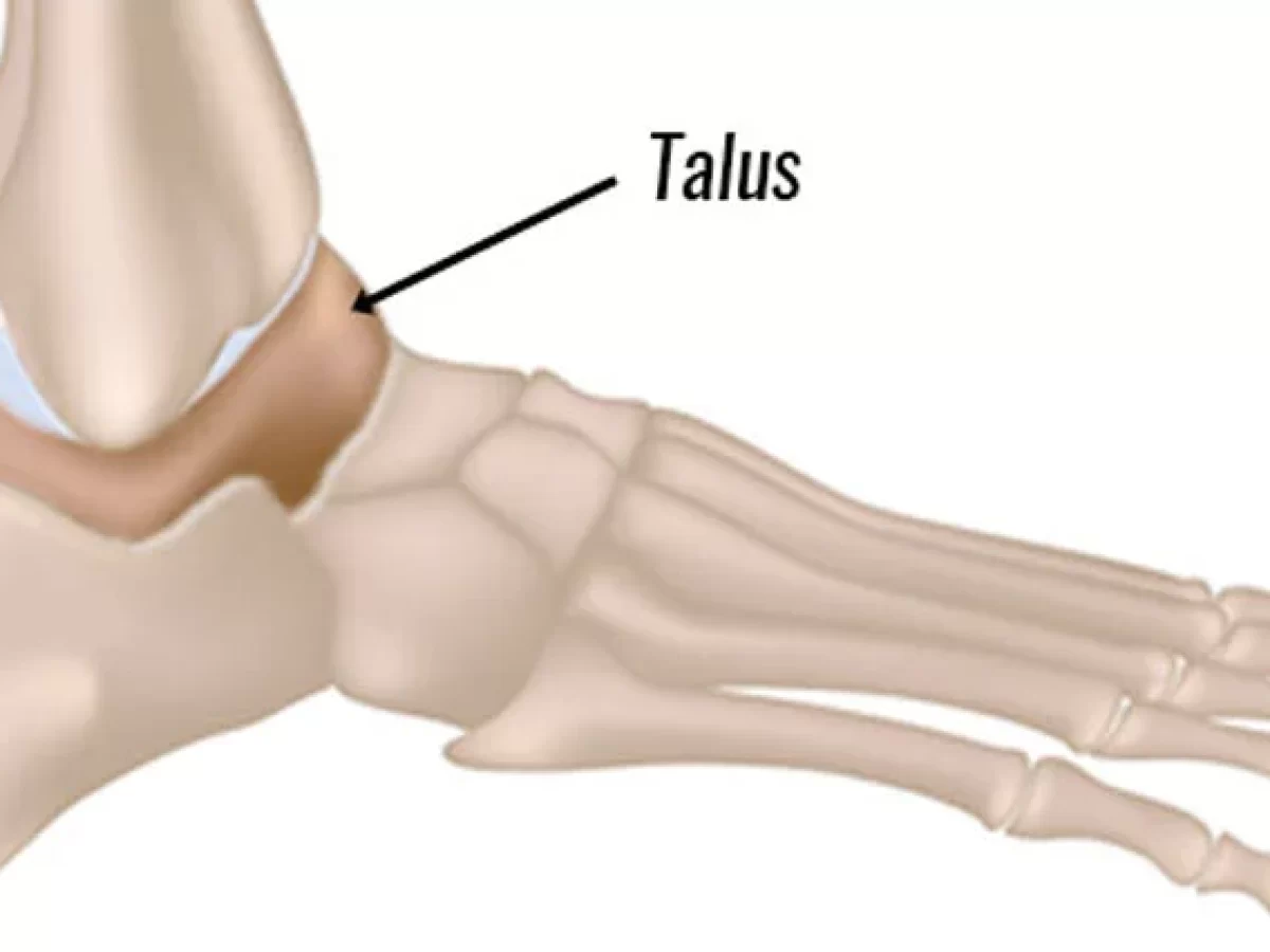
The talus bone, also known as the ankle bone, is a critical structure in the human foot that plays a pivotal role in mobility and weight-bearing.
The talus bone’s typical form allows it to align with the shin and calf bones, the tibia, and the fibula, to comprise the ankle joint.
It facilitates fluid ankle movement and supports your weight. The talus is more susceptible to complications than other bones, and injuries and damage to it may take longer to recover.
What is Talus bone?
- Your ankle endures a small-sized bone known as the talus bone. The astragalus bone is another name for it, which joins the calf (fibula) and shin (tibia), and makes up the joint that links the ankles.
- The talus, or ankle, is critical for the ankle joint’s strength and flexibility. Your capacity to stand and move is greatly influenced by the talus, despite its small shape. It balances your weight and allows for elegant ankle movement.
- It is in charge of transferring weight from the leg to the foot and allowing movements like plantarflexion, which points the foot downward, and dorsiflexion, which raises the foot upward.
Anatomy of Talus Bone
The talus has a saddle-like form. It creates a domed bulge in the midpoint and two widened lower ends. Your ankle moves more smoothly because of the layer of cartilage covering it, which serves as a cushion, shock absorber, and lubricant. The talus has no muscle attachments, different from many other bones.
There are three sections to the talus:
- Talus head: It is revealed on the medial side of the bone. At the rear of your foot, the head connects to your navicular bone. When the navicular and cylindrical bones articulate, the talonavicular joint takes shape.
- Talus body: The majority of the weight is supported by the talus body, which makes up the bulk of the bone when walking or running. It forms the ankle joint by articulating with the tibia on its convex superior surface. The concave inferior surface joins the calcaneus bone to form the subtalar joint.
- Talus neck: The phrase “talus neck” refers to the part of the bone that links to the talus head and body. It bends in the direction of the inside of your foot.
Articulation Surface
Numerous articular surfaces on the talus bone enable a range of motions and interactions with other foot and ankle bones.
- The tibia and fibula come together superiorly via the talar arch to form the ankle’s transverse joint.
- The calcaneus meets with a broad, concave oblique facet Inferoposteriorly establishing the talocalcaneal joint.
- Two facets that articulate with the calcaneus to create a portion of the talocalcaneonavicular joint are located anteroinferiorly.
- Navicular bone (circular notch on the posterior surface) and talar head (domed articular surface).
Muscles
- Not having any muscular attachments to the talus is important because it indicates that the talus lacks supplementary blood supply sources.
- There is a comparatively significant chance of developing post-traumatic, avascular necrosis if the talus has a vascular compromise due to traumatic injuries, such as displaced fractures.
Ligament
- The anterior talofibular ligament
- The posterior talofibular ligament
- Talocalcaneal ligaments
- Tarsal sinus ligaments.
- Cervical ligament
- Talocalcaneal Interosseous Ligament
- The deltoid ligament
- The anterior tibiotalar ligament
- Posterior superficial tibial ligament
- The posterior deep tibiotalar ligament
- The dorsal talonavicular ligament
Blood supply
- This artery unites the posterior tibial area to the sinuses and the medial side of the body.
- The head and neck receive blood via the anterior tibial and dorsal pedis arteries.
- The lateral region of the body and the sinuses receive their blood supply from the peroneal artery.
Nerve supply
- The deep peroneal nerve
- The tibial nerve
- The saphenous nerve
- Sural nerves
The function of the Talus Bone
The talus bone, commonly known as the ankle bone, is crucial in the foot and performs various critical roles.
- Your ankle joint’s formation.
- Keeping your leg’s weight supported.
- Putting your foot up and down.
- By shifting the rear of your foot from side to side, you can keep your balance.
- Stabilizing your foot’s arch.
- Supporting bones in your foot, heel, and ankle.
Conditions and Disorders of the Talus Bone
Talus fractures
Your talus may be broken by incidents like falling or car crashes. Talus fractures can also occur during athletic activities. The symptoms of a fracture are:
- Pain.
- Swelling.
- Tenderness.
- Having difficulty walking.
- Bruise or discoloration.
- A deformity or protrusion that is abnormal for the body.
If you are worried that you may have been wounded or have a fracture, go to the emergency division fast.
Osteoporosis
Osteoporosis weakens the bone structure, enhancing the possibility of sudden fractures. Numerous individuals are unknowing that they have osteoporosis until a bone ruptures There are generally no noticeable signs. Women, those your provider about a bone density screening, which can detect osteoporosis before it causes a fracture.
Other typical conditions that might influence your talus are:
- Arthritis of the feet and ankles.
- Tarsal tunnel syndrome.
- Osteonecrosis (avascular necrosis).
Diagnosis of the Talus Bone
The primary method used to figure out the condition of your bones is to get a bone density test. The terms DXA and DEXA scan are frequently used to describe it. Low-dose X-rays are used in a bone density test for bone strength. It’s a way to quantify bone loss with aging. If you’ve been injured, your physician or surgeon may recommend imaging testing, such as:
- X-rays.
- Magnetic resonance imaging (MRI).
- A CT scan.
Treatment for the Talus Bone
Unless you have a fracture or another ankle injury, your talus usually does not require treatment. You may require therapy if you have been diagnosed with osteoporosis.
Talus fracture treatment
- The type and cause of your fracture will determine how it is treated. To heal, you may require immobilization, such as a splint or cast, and even surgery to realign and fix your bone in its proper position.
Osteoporosis Treatment
- Vitamin and mineral supplements, medications, and physical activity are all alternatives for treating osteoporosis.
- Exercise and supplementation are typically the only ways to avoid osteoporosis. A personalized treatment plan that examines your specific requirements and bone health will be developed in coordination with your doctor.
Maintain your Talus Bone healthy
Keep your talus bone in good condition.
You can preserve the health of your bones and your body by sticking to a nutritious diet, exercising regularly, and seeing your doctor regularly. If you are over 50 or have a family history of osteoporosis, consult your doctor about a bone density scan. Obey these known safety precautions to lessen the danger of injury:
- Wear a seatbelt at all times.
- Sport the suitable protection gear for all sports and activities.
- Make sure your home and workplace are free of hazards that might fall on you or others.
- To gain something at the house, always utilize the right devices or equipment. Refrain from standing on desktops, benches, or chairs.
- Follow an exercise and nutrition plan that will promote bone health.
- Use a walker or cane if you have problems walking or are at greater risk of falling.
Summary
Your capacity to walk, stand, and move is greatly influenced by the talus, a little bone. See your doctor as soon as you experience any pain or other symptoms in your ankle because it may take longer to mend and has a higher chance of complications than other bones.
If you have been traumatized, get to the emergency hospital immediately. Discuss your risk of osteoporosis with your healthcare practitioner and find out how you can maintain your bone health as you age.
FAQs
What is the bone called the talus?
The talus bone is a little bone located in your ankle. The astragalus bone is another name for it. The second greatest bone in the hindfoot, or rear of the foot, is the talus. The only larger bone is the calcaneus (heel).
With a shattered talus, is it possible to walk?
It won’t support any weight or allow you to walk on it. For eight to twelve weeks or more, you might be necessary to wear an immobile plaster cast, based on how serious your injury is. The length of time your foot is in a cast increases with the severity of your injury. Even after the bone heals, you can continue to experience pain, stiffness, and swelling.
How is the talus bone fixed?
Due to the high-intensity impact that produces the damage, many talus fractures require surgery. However, stable, well-aligned fractures frequently respond well to non-surgical treatment. This is typically accomplished through the combination of therapy with immobilization.
References:
- Professional, C. C. M. (2024a, May 1). Talus Bone. Cleveland Clinic. https://my.clevelandclinic.org/health/body/23416-talus-bone
- Wikipedia contributors. (2024, August 14). Talus bone. Wikipedia. https://en.wikipedia.org/wiki/Talus_bone
- Hegazy, M. A., Khairy, H. M., Hegazy, A. A., Sebaei, M. a. E. F., & Sadek, S. I. (2023). Talus bone: normal anatomy, anatomical variations, and clinical correlations. Anatomical Science International, 98(3), 391–406. https://doi.org/10.1007/s12565-023-00712-y
- Patel, D. (2023a, July 9). Talus Bone – Anatomy, Function & Related Conditions. Samarpan Physiotherapy Clinic. https://samarpanphysioclinic.com/talus-bone/

One Comment