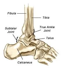
Subtalar Joint
Introduction
The subtalar joint, located just below the ankle joint, plays a critical role in foot and ankle biomechanics. It connects the talus (a bone in the foot) with the calcaneus (heel bone), enabling the complex movements of inversion and eversion, which allow the foot to adapt to uneven surfaces.
As a synovial joint, this allows smooth gliding and controlled motion. This joint is referred to in the literature as the anatomical subtalar joint and the subtalar joint proper.
Subtalar (posterior) and talocalcaneonavicular (anterior) joints are anatomical units that are considered functional units in clinical practice. This functional region’s principal anatomical features include the tarsal sinus, tarsal canal, and acuminate interosseous tunnel.
The anatomical subtalar joint is a single synovial articulation that is formed by the convex posterior articular surface of the calcaneus and the posterior calcaneal articular aspect of the talus. To hold bones together, there are ligaments, capsules, and interosseous connections.
This joint makes supination and pronation, the two essential synthesis movements. There is considerable movement in ligaments, tendinous tissues, and joints.
Both eversion and inversion permit:
- To rotate or alter direction, use your foot and ankle.
- Maintain equilibrium
- Controlling movement across uneven ground.
- Serve as a cushion for impact.
Pathology
The majority of forms of arthritis affect the subtalar joint, especially if it has undergone prior sprains or fractures with those to the talus or calcaneum. The typical signs of subtalar joint arthritis involve difficulties walking on uneven surfaces and a limited range of motion in the joints. In flat feet, the joint usually appears more horizontally.
Biomechanics
The convex posterior of the talus and calcaneus establishes the movement and rotation axis complex of the subtalar joint. Manter states that a 10° circle rotation results in a 1.5 mm talus movement. Translation, rotation, or screw-like movement were noted during Inman’s skeletal research. It’s possible that the approach—which involved both in vivo studies and static radiography images—led to the significant degree of flexibility detected in the subtalar joint’s movement axis.
The subtalar joint can move in two directions: five to ten degrees inversion and twenty to thirty degrees inversion. Ankle joint position and various procedures can assist in explaining the wide variety in range.
Tendons support the ankle during stride and stance; depending on the position of the subtalar joint, evertors, and invertors require the tibialis posterior and peroneus longus.
Anatomy
To form the subtalar joint, these two components come together. The anterior talar head is located on the anterior and middle sides of the calcaneus and is stabilized by the navicular bone and acetabulum pedis.
The interosseous calcaneal ligament divides and separates the larger posterior talus area. There are variants in anatomy for the anterior and center surfaces: 42% have an oblong shape, 22% have a “bean” form, and 36% have a full separation.
The subtalar joint may be harmed by both intrinsic and extrinsic ligaments, among them the cervical ligament and the interosseous talocalcaneal ligament. Anterolateral rotation that is not normal might result from interosseous ligament dysfunction and cause instability. Studies show increased subtalar movement and instability, but the significance of the calcaneofibular ligament for subtalar stabilization is different. Movement and stability of the subtalar joint are also facilitated by the inferior extensor retinaculum.
Structure
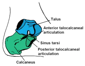
The Multiarticular ST joint is composed of three articulated facets that form a gliding surface. To make up the ST joint, there are two parts:
At a somewhat oblique angle, the talus is situated on the front side of the calcaneus.
Between the two bones, there are three areas of articulation: one posterior and two anterior. Together, these three articulations are known as the anterior, middle, and back aspects.
- The calcaneus muscles and the talar head articulate at the anterior subtalar joint. This allows forward sliding, or anterior articulation.
- Motion from side to side is enabled by the medial subtalar joint.
- The posterior subtalar joint is formed by the concave surface of the talus and the convex posterior section of the calcaneus coming together. which allows posterior articulation, or moving backward.
Although the anterior facet’s articulation and lateral alignment with the talus head, the middle aspect’s floor is given by the sustain tenaculum tali. The center and anterior faces of certain people are related, leading to only one articulation. The tarsal canal divides the three facet bones into those that are largest and greatest in size.
Location
The heel, ankle, and hindfoot develop from the seven tarsal bones that compose each foot. Calcaneus, Talus, and Proximal Tarsal are the bones next to the ankle. The Achilles tendon is connected to the calcaneus, the largest and strongest bone.
The talus, or superior tarsal bone, is the bone that has connections to the tibia and ankle. Three subtalar facets and layers are situated in the groove above the calcaneus bone’s front part. It is the interosseous talocalcaneal ligament separating the calcaneus and talus.
Stability
In the subtalar joint are the synovial membrane, the fibrous layer, three ligaments, and the cap.
- Posterior talocalcaneal ligament
- Medial talocalcaneal ligament
- Lateral talocalcaneal ligament
Strength and stability are provided to the sinus tarsi cavity by the interosseous talocalcaneal ligament, which joins the talus and calcaneus.
Articular surfaces
The synovial articulation known as the anatomic subtalar joint joins the talus and calcaneus. It has two articular facets that are covered in hyaline cartilage. Two different articular surfaces consist of this joint: the concave posterior calcaneal aspect on the inferior surface of the talus and the convex posterior talar articular surface on the superior aspect of the calcaneus.
Ligaments and joint capsule
Single, loose joint capsules around the subtalar joint. The synovial membrane, as in other synovial joints, envelops the joint capsule. The lateral, medial, and posterior ligaments of the joint capsule are the main ligaments that establish joint movement. The articular surfaces, the floor of the tarsal sinus, and the roof make the subtalar joint capsules. In the tarsal sinus, the calcaneus and talus join. Situated directly in front of the subtalar joint, it is a small transmit.
The talocalcaneonavicular and subtalar joint capsules inside the tarsal sinus evolved into the talocalcaneal interosseous ligament.
The calcaneofibular ligament, lateral talar process, and lateral calcaneal surface are connected by an oblique ligament called the lateral talocalcaneal ligament. It is a flat fibrous group. It is situated far below the ligament that connects the fibula and calcaneus.
A portion of its distal sarcophagi connects with the ankle joint’s medial deltoid ligament.
The calcaneus and the ankle are connected by the posterior talocalcaneal ligament. The two robust, thick fibrous units that comprise the interosseous talocalcaneal ligament are located in the tarsal sinus. This deep extension of the inferior extensor retinaculum forms the interosseous ligament group this joins.
This ligament, which connects the talocalcaneal and talocalcaneonavicular joints, covers the central position that controls both joints’ function. The subtalar ligament’s primary role is to stabilize the joint during both active and passive motion. Foot mobility is restricted by the rigidity of the interosseous talocalcaneal ligament.
Horizontal direction characterizes both the tarsal sinus and the anterior talocalcaneal ligament. The calcaneum’s upper surface, also known as the talar neck, is where the tubercle ends. Additionally, with pronation, this ligament is stiff.
Innervation
The medial or lateral plantar nerves feed the plantar portion of the subtalar joint, while the deep fibular nerve governs the dorsal side.
Neurovascular Supply
Two arteries and two nerves provide blood to the subtalar joint. The major arteries that supply blood are the fibular and posterior tibial arteries.
The dorsal side of the joint is innervated by the deep fibular nerve, whereas the plantar section is innervated by the lateral or medial plantar nerve.
Blood supply
The fibular artery and the posterior tibial artery deliver blood to the subtalar joint.
Function of Subtalar Joint
The joint permits the foot to invert and evert, even though the plantarflexion or dorsiflexion only partially recovers. It is thought that the middle aspect may be the location of the subtalar joint’s center of rotation.
These joints are also known as gliding joints or plane synovial joints. Between the calcaneus and talus, it provides a stable connection. The tilt from horizontal has changed significantly.
Subtalar joint components involve the talocalcaneal and talocalcaneonavicular joints. The former was originally described as an anatomic subtalar joint. This represents the subtalar joint’s typical range of motion. The midfoot can pronate and supinate due to joint articulations.
Movement
The results of rotation and gliding at the subtalar joint are pronation and supination. The talocalcaneonavicular and calcaneocuboid joints provide much of the foot’s mobility.
From the calcaneus tubercle to the talus neck, supination and pronation happen on a single axis with an average rotation angle of 42° upward and anterior to the transverse plane.
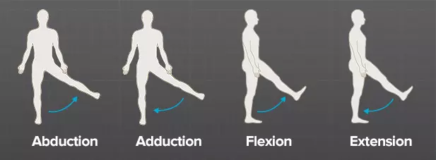
Pronation results from eversion, abduction, and dorsiflexion at joints, whereas supination is characterized by inversion, adduction, and plantarflexion. Primary movements ought to occur by coincidence and never alone.
The two main axes for movement for the transverse tarsal and subtalar joints are eversion and inversion. The ranges of these motions vary according to the origin; they range from 5° to 10° for eversion and from 25° to 30° for inversion.
The lateral side of the foot appears inferior when the foot is everted, or inverted, and the sole bends inward toward the midline. Abduction and adduction centers are located on vertical axes in the transverse plane. The range of motion for adduction and abduction is quite similar to that for inversion and eversion, but it depends on the particular purpose of the axis of rotation. More adduction/abduction and less RoM for inversion/eversion happen at an inclination of less than 42°, although the opposite occurs nearer the leg’s long axis.
Abduction/adduction and inversion/eversion are analogous in these little parallel motions.
Muscles acting on the subtalar joint
The extrinsic muscles that extend the toes—the extensor digitorum logus and the extensor hallucis longus—assist the fibularis longus, fibularis brevis, and fibularis tertius in producing pronation.
Clinical Significance
Injuries like sprains or fractures, as well as illnesses like arthritis or instability, can cause issues with the subtalar joint. Pain, restricted range of motion, and trouble walking or running can result from subtalar joint problems. Conservative measures including rest, physical therapy, orthotics, or, in extreme situations, surgery, may be part of the treatment choices.
For a proper diagnosis and treatment of any subtalar joint problems, it is imperative to seek advice from a skilled orthopedic specialist or healthcare practitioner.
A painful condition known as sinus tarsi syndrome can affect athletes. Subtalar joint synovitis and enduring ankle pain are among its adverse effects. After a physical examination is used to make the diagnosis, treatment consists of physical therapy and anti-inflammatory medications.
Gout
Gout is a type of arthritis that primarily affects the first metatarsophalangeal joint or big toe, but it can also cause inflammation and pain in the subtalar joint.
Juvenile idiopathic arthritis
The subtalar joint is frequently the first joint to be affected by this kind of pediatric arthritis, which has an unknown cause.
Subtalar instability
A weakness in the lateral direction where the ankle can suddenly “give way.”The severe strain on the ligament may result in persistent ankle inflammation and recurrent ankle twisting.
Subtalar dislocation
This injury, sometimes known as “basketball foot,” usually happens when you land hard on the inside or outside of your foot.
Summary
The heel is made up of the talus and calcaneus bones, which join the other tarsal bones of the hindfoot at the subtalar joint.
The major function of the subtalar joint is to facilitate walking. Acute traumatic injuries such as subtalar dislocation and chronic disorders like osteoarthritis that result from wear and tear can occur because of the consistent and repeated stress exerted on them.
In a “crush” type of injury, the calcaneus is frequently shattered. The talus is usually forced into the calcaneus when someone falls onto their heel from a height. A comminuted fracture occurs when a bone fractures into many fragments. A calcaneus will appear shorter and wider.
Even with treatment, persistent issues may arise from a calcaneal fracture. Damage to the subtalar joint is often the cause of arthritis. Walking on uneven ground can be very uncomfortable for the patient since they experience pain during inversion and eversion.
FAQs
Which joint is this, the ankle or the subtalar joint?
The calcaneus and talus are involved in the subtalar joint, also referred to as the talocalcaneal.
What does the term “subtalar” mean?
Above the suprasellar joint is the talus, or ankle bone.
What is the purpose of the subtalar joint?
The calcaneus and talus, two tarsal bones of the foot, become linked by the subtalar (ST) joint. The synovial joint known as the St joint facilitates easier rotation, everting, and inversion of the foot.
Which muscles provide subtalar joint stability?
The lateral ankle stabilizers and major investors Peroneus longus, brevis, and Tertius are essential for pronator and supinator action.
What neutral position does the subtalar joint have?
Physicians use the STJoint neutral position for cast representations in a biomechanical functional orthosis, but there is no exact or standardized approach.
What are the subtalar joint’s tendons?
The digitorum longus, hallucis longus, and tibialis posterior tendons are supported by the sustentaculum tali, part of the middle calcaneal aspect.
Is the subtalar joint a nerve?
The subtalar joint receives its supplies from two arteries and two nerves. The posterior tibial and fibular arteries are the blood vessels that supply blood. The dorsal side of the joint is supplied by the deep fibular nerve, whereas the plantar facet is provided by the lateral or medial plantar nerve.
What three components make up the subtalar joint?
Subtalar joints are intricate tri-planar joints that result from the fusion of the calcaneus’ superior surface and the talus’ inferior surface. For the joint connecting the two tarsals, the anterior, middle, and posterior aspects are the three points of articulation.
Which muscles are inverted by the subtalar joint?
The function of the tendons depends on how they adhere to the subtalar joint axis. The evertors involve the extensor halluces longus, extensor digitorum longus, and peroneus longus/brevis. Associated with inversion are the flexor digitorum longus, flexor hallucis longus, flexor posterior, and tibialis anterior muscles.
How do you evaluate subtalar joints?
An inclinometer, also called an angle finder, has been proposed as a tool for assessing STJ position in a weight-bearing (closed kinetic chain) posture. A different method for figuring out the location and mobility of the STJ is to assess the navicular tuberosity’s weight-bearing height.
What are the subtalar joint injuries?
Sprains: These injuries cause stretches or tears in the ligaments. Strains: These injuries cause strained or torn tendons or muscles. Fractures: The American Academy of Orthopaedic Surgeons says that pain in the subtalar joint may be caused by broken talus bones. Dislocation: This kind of damage is characterized by the popping or slipping of bones out of place.
What additional terms do you know for the subtalar joint?
A connection exists between the calcaneus and talus and the subtalar joint, also known as the anatomical subtalar joint.
References
- Gameti, D. (2023, July 31). Subtalar Joint – Anatomy, Function, Movement. Samarpan Physiotherapy Clinic. https://samarpanphysioclinic.com/subtalar-joint/
- Wikipedia contributors. (2024, February 11). Subtalar joint. Wikipedia. https://en.wikipedia.org/wiki/Subtalar_joint
- Dpm, C. M. (2022, September 11). The Anatomy of the Subtalar Joint. Verywell Health. https://www.verywellhealth.com/what-is-the-subtalar-joint-1337686

One Comment