Metatarsal Bones
Introduction
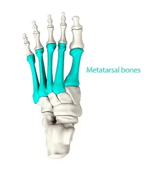
The metatarsals are the cylinder-shaped bones that make up the foot’s core. They are designated numerically and begin from the medial side outward. Letters one through five correspond to the metatarsal bones.
The phalanges and tarsals are split by these bones. The bases of every single bone are going to relocate in tandem with at least one tarsal bone in the tarsometatarsal joint area. At the metatarsophalangeal joint, the metatarsal bones communicate to the phalanges, or toe bones. The metatarsals are long bones with a convex form (arch upward) that give the foot its arch. To enable movement in the foot, they work with ligaments, tendons, and connective tissues.
The metatarsal and tarsal bones comprise the foot’s major arches, which support weight-bearing and walking. Misuse or overuse of these bones can cause them to fracture, strain, or inflame. Foot immobilization (e.g., casting) can assist in healing metatarsal fractures and sprains.
What are Metatarsal Bones?
Five long bones described as the metatarsal bones, metatarsus, are located in the center of the foot, between the phalanges and the tarsal bones, ankle, and heel also referred to as metatarsi. The first, second, third, fourth, and fifth metatarsal bones are designated by numbers from the medial side, or great toe side, and are commonly displayed with numerals in Roman characters.
Anatomy of the Metatarsal Bones
Structure
- The five metatarsals are prolonged, dorsal curved bones that approximate a head on the distal end, a base on the proximal end, and a shaft or body. Prismoidal in shape, the body curves longitudinally to be concave below and somewhat convex above, tapering progressively from the tarsal to the phalangeal extremities. The base, also known as the posterior extremity, is a rectangular wedges component that communicates with the adjacent metatarsal bones throughout its sides alongside the tarsal bones proximally.
- Its dorsal and plantar surfaces are roughly designed to assist in ligament attachment. The convex articular surface of the skull or distal extremity is rectangular from above downward and extends more backward below than above. Each collapsed side has a tubercle on top of it and an aperture for ligamentous attachment. An articular eminence that is continuous with the terminal articular surface marks each side of its plantar surface, with an anteroposterior groove for the flexor tendons to pass through.
- The first metatarsal positions itself proximally during growth and development, whereas the other growth plates are situated distally on the metatarsals. However, an additional growth plate on the distal first metatarsal is rather frequent.
First Metatarsal
The foot’s first metatarsal is its smallest and most viscous portion. The base frequently lacks facets, but on occasion one develops laterally where it articulates with the second metatarsal. It uses the medial cuneiform to define it at the base, distally.
A tuberosity can be seen medially here. The prism-shaped shaft is robust. Two distal grooved areas on the plantar surface are where the two (hallucal) sesamoid bones converge. Human hallucal sesamoids are consistently located and visible on the heel side of the first metatarsal head. The shaft and the first proximal phalanx articulate distally.
The following are the first metatarsal’s muscle attachments:
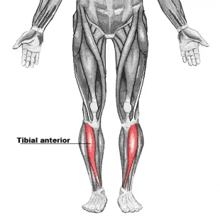
- The base of the fifth metatarsal is where the tibialis anterior and fibularis longus muscles originate.
- Tuberosity: the muscle of the fibularis longus.
- External positioning characterizes the first dorsal interosseus muscle.
Second Metatarsal
The longest metatarsal, the second one, has four unique characteristics at the base of it. They engage in interaction with the third metatarsal as well as the medial, middle, and lateral cuneiforms. The first metatarsal and the medial region of the base are not well connected. It forms a line with the second proximal phalanx on the distal side.
The following are the second metatarsal’s muscle attachments:
- First dorsal interosseus muscle, medial shaft.
- The second dorsal interosseus muscle of the lateral shaft.
Third metatarsal
The lateral cuneiform attaches to the third metatarsal’s triangular base proximally. It shares an exterior with the second metatarsal via two facets in the middle and with the fourth metatarsal via one facet externally. Additionally, the third terminal phalanx unites the head.
The following are the third metatarsal’s muscle attachments:
- First plantar interosseus and second dorsal interosseus muscles in the medial shaft.
- The third dorsal interosseus muscle of the lateral shaft.
Fourth Metatarsal
The base of the fourth metatarsal is more constrained than the third, even though it has three joint facets. At its proximal end, a quadrilateral facet connects it to the cuboid. The third metatarsal aligns with an oval facet medially, while the fifth connects with a single facet on the lateral side. At a particular length, the head connects with the fourth proximal phalanx.
The following are the fourth metatarsal’s muscle attachments:
- Third dorsal interosseus and second plantar interosseus muscles in the medial shaft.
- The fourth dorsal interosseus muscle of the lateral shaft.
Fifth Metatarsal
The side border of the foot reflects and feels the tuberosity that extends laterally to the base of the fifth metatarsal. Through a triangular surface, the base of the fifth metatarsal joins proximally with the cuboid and inwardly with the fourth metatarsal. The fifth proximal phalanx also outlines with its head.
The following are the fifth metatarsal’s muscle attachments:
- Peroneus tertius muscle: dorsal base.
- Tuberosity: muscle of the fibularis brevis.
- Included in the flexor digiti minimi brevis of the plantar base is this muscle.
- Third plantar interosseus and fourth dorsal interosseus muscles in the medial shaft.
Articulation
Tarsometatarsal joints
The metatarsals serve as a link between the toes and the ankle. They are categorized as I through V from medially to laterally based on examinations made from the front aspect of the foot. One or more distal tarsal bones, specifically the cuboid and cuneiform bones, articulate with the proximal base. The term “tarsometatarsal joints” describes these articulations.
Metatarsophalangeal joints
The metatarsophalangeal joints are produced when their distal heads come into contact with the adjacent proximal phalanx. Two sesamoid bones can additionally determine the head of the first metatarsal on the plantar province of the foot.
Intermetatarsal joints
Additionally, when the bases of the metatarsals interact, intermetatarsal joints take shape. The perfect places in the metatarsals that interact with the adjoining bones are identified as articular facets.
Arches of the Foot
- A key component of the foot’s three arches is the metatarsals. The bones that comprise the arch of the foot structure are as follows:
- The medial longitudinal arch is assembled from the up of the talus, calcaneus, navicular, all three cuneiforms, and metatarsals one through three.
- Metatarsals four and five, the cuboid, and the calcaneus are all part of the lateral longitudinal arch.
- The transverse arch is assembled from the bases of the cuboid, cuneiforms one and three, and all five of the metatarsals.
Function of the Metatarsal Bones
The metatarsals work in tandem with the calcaneus to help distribute the body’s weight. The metatarsus and the ground make touch with each other at five main places.
- The Frist metatarsal head and two sesamoid bones
- The second metatarsal head
- The third metatarsal head
- The fourth metatarsal head
- The fifth metatarsal head
Relevant Condition
Metatarsalgia
Discomfort in the forefoot regarding one or more metatarsal heads can be described as metatarsalgia. Primarily transmitted by the metatarsals’ physical features, which influence how they relate to each other and the remaining bones of the foot.
Fracture
Although they are rare, metatarsal fractures happen when a heavy object rolls or falls on the foot. Ballet dancers who lose their balance while on their toes may also sustain these fractures.Because the metatarsals bear the whole weight of the body, one or more of them may break.
Gout
Gout is an inflammatory disease that produces high blood levels of uric acid and crystal deposits in the joints and surrounding tissues. The metatarsophalangeal joint of the great toe is often the first to be affected by gout. This joint may become sore and swollen due to gout but when this joint is afflicted, it becomes known as podagra. This joint may also be quite painful due to osteoarthritis.
Valgus Hallux
Hallux valgus is a foot deformation that explains a laterally replaced great toe (hallux) and a medial deviation of the first metatarsal. It is frequently caused by pressure from shoes or degenerative joint conditions. Hallux valgus, which is more frequent in females, results in a medial displacement of the first metatarsal and a lateral shift of the sesamoid bones. Consequently, the first and second metatarsal heads remain separated by the sesamoid bones. A subcutaneous bursa may develop as a result of swelling in the surrounding tissues. This bursa can be extremely painful when it becomes inflammatory.
FAQs
Why do my metatarsals hurt?
The ball of your foot gets painful and swollen when you have metatarsalgia (pronounced “met-uh-tahr-SAL-juh”). You may develop it if you engage in activities that require running and leaping. There are additional causes, such as foot abnormalities and shoes that are too tight or loose.
Can I walk with a broken metatarsal bone?
Walking with a fractured foot will be painful. You may be able to walk lightly with a 5th metatarsal fracture, but it requires immobilization to heal correctly.
What is the metatarsal bone?
The term metatarsals refers to the five long bones that make up a single foot. Numerals I through V, medial to lateral, are used to identify them.
To what extent is a metatarsal fracture severe?
If adequately treated, metatarsal fractures are supposed to recover on their own. If not identified and treated, some of the following problems may occur: A metatarsal stress fracture can worsen over time if the bone is subjected to repeated stress.
What is the treatment for metatarsals?
Elevate your foot after standing or walking. You may need to give up your favorite sport for a spell, but you may stay active with low-impact exercises like swimming or cycling. Several bits a day, employ cold packs on the affected spot for 20 minutes at a duration.
References
- Wikipedia contributors. (2024c, August 27). Metatarsal bones. Wikipedia. https://en.wikipedia.org/wiki/Metatarsal_bones#:~:text=The%20metatarsal%20bones%20or%20metatarsus,and%20the%20phalanges%20(toes).
- Metatarsal bones. (2023, October 30). Kenhub. https://www.kenhub.com/en/library/anatomy/metatarsal-bones
- TeachMeAnatomy. (2023, July 16). Bones of the Foot – Tarsals – Metatarsals – Phalanges – TeachMeAnatomy. https://teachmeanatomy.info/lower-limb/bones/bones-of-the-foot-tarsals-metatarsals-and-phalanges/
- The Healthline Editorial Team. (2018b, January 21). Metatarsals. Healthline. https://www.healthline.com/human-body-maps/metatarsal-bones
- The Editors of Encyclopaedia Britannica. (2024b, December 18). Metatarsal | Anatomy, Structure, & Function. Encyclopedia Britannica. https://www.britannica.com/science/metatarsal

3 Comments