Interphalangeal Joints of the Foot
The interphalangeal (IP) joints of the foot are the articulations between the phalanges (toe bones). These hinge joints allow for flexion and extension, contributing to toe movement and balance.
The toes’ interphalangeal joints are synovial. The proximal and distal interphalangeal joints on the four lesser toes connect three phalanges, while the hallux has a single IP joint that connects two phalanges.
Introduction
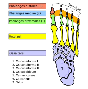
Interphalangeal joints are the points of articulation between the foot’s successive phalanges. Two of the four lateral toes and one on the big toe make up each foot’s nine interphalangeal joints. Frequently, they can be divided into:
- Between the proximal phalanges’ heads and the middle phalanges’ bases are the proximal interphalangeal joints.
- Between the bases of the distal phalanges and the heads of the middle phalanges are the distal interphalangeal joints.
The big toe has a single interphalangeal joint between the proximal and distal phalanges because it lacks a middle phalanx.
The flexion (plantarflexion) and extension (dorsiflexion) of the middle and distal phalanges are examples of the movement along a single axis that is possible with uniaxial hinge joints, a kind of synovial joint that includes the foot’s interphalangeal joints.
The articulations between neighboring phalanges in the foot are called interphalangeal joints. Each foot has nine interphalangeal joints. The lesser digits each have a proximal and a distal interphalangeal joint, whereas the hallux has a single interphalangeal joint.
Since the great toe only has two phalanx bones (proximal and distal phalanges), it only has one interphalangeal joint, or “IP joint” as it is commonly shortened. A proximal interphalangeal joint (abbreviated “PIP joint”) connects the proximal and middle phalanges, while a distal interphalangeal joint (abbreviated “DIP joint”) connects the middle and distal phalanges.
A plantar (underside) and two collateral ligaments are present in every interphalangeal joint, which is a ginglymoid (hinge) joint. The dorsal ligament locations in this ligament arrangement are supplied by extensor tendons, which are comparable to the metatarsophalangeal articulations.
Articular surfaces
The phalanges of the toes articulate with one another to form the foot’s interphalangeal joints. A proximal and distal set of interphalangeal joints can be found in toes 2–5. The concave articular surface on the neighboring base of a middle phalanx and the convex trochlear surface on the head of a proximal phalanx produce a proximal interphalangeal joint.
A homologous process forms the distal interphalangeal joints of toes 2–5, between the concave articular surface on the neighboring bases of a distal phalanx and the trochlear surfaces on the heads of a middle phalanx.
As previously stated, the trochlear surface of the proximal phalanx’s head and the concave articular surface of the distal phalanx’s base form the big toe’s sole interphalangeal joint.
Joint capsule
A joint capsule lined with synovial membrane entirely encloses each interphalangeal joint. Attached to the articular borders, the joint capsule is strengthened by the plantar ligament, collateral ligaments, and the extensor expansions of the foot’s intrinsic and extrinsic muscles.
Ligaments
The proximal and distal interphalangeal joints are supported and stabilized by two different kinds of ligaments:
- Strong ligaments called collateral interphalangeal ligaments are located on the medial and lateral sides of each joint. They run medially and proximally from the tiny tubercles on either side of the proximal phalanx’s head to the base of the following phalanx.
- The plantar surface of the phalangeal heads at the interphalangeal joints contains a dense fibrocartilaginous plate called the plantar interphalangeal ligament, which is not a real ligament.
Innervation
The medial and lateral plantar branches of the tibial nerve’s appropriate plantar branches supply nerves to the interphalangeal joints.
Dorsal digital branches, which originate from the deep fibular (peroneal), intermediate dorsal cutaneous, and sural nerves, provide minor innervation to the lateral toes’ interphalangeal joints. Additional innervation of the big toe’s interphalangeal joint is provided by the superficial fibular nerve’s medial dorsal cutaneous branch.
Blood supply
Through an anastomosis created by the lateral and deep plantar arteries, digital branches of the plantar arch provide arterial blood supply to the foot’s interphalangeal joints.
Movements
The only motions allowed in the interphalangeal joints, which are uniaxial hinge joints, are flexion (plantarflexion) and extension (dorsiflexion), which take place in the sagittal plane around a frontal axis. Extension is restricted by the plantar and collateral ligaments, but flexion is possible to a significant degree. Additionally, the proximal interphalangeal joints exhibit noticeably more movement than the distal ones.
The interphalangeal joints are fully extended in the close-packed position and slightly flexed in the loose-packed (or resting) position. Flexion is more restricted than extension in these joints’ capsular pattern, which is the loss of passive range of motion during inflammation.
Muscles acting on the interphalangeal joints
The main flexor of the distal interphalangeal joint is the flexor digitorum brevis, whereas the flexor digitorum longus is the primary flexor of the proximal interphalangeal joint. Flexor hallucis longus is responsible for flexing the big toe’s interphalangeal joint.
The lubricants, interossei, extensor digitorum longus, and extensor digitorum brevis are the main extensors of the interphalangeal joints.
Clinical significances
The articulations between the phalanges (toe bones) make up the foot’s interphalangeal (IP) joints. The big toe, which has two phalanges rather than three, has only one interphalangeal joint. All other toes have one distal interphalangeal (DIP) joint. When walking, these joints are essential for weight distribution, balance, and mobility.
1. Osteoarthritis (OA)
- Pain, stiffness, and a decreased range of motion can result from degenerative changes in the IP joints.
- More prevalent in elderly people or people with a history of trauma.
2. Rheumatoid Arthritis (RA) and Other Inflammatory Conditions
- Synovitis, joint abnormalities, and erosion of the IP joints can be brought on by psoriatic arthritis, RA, and other inflammatory diseases.
- Causes functional impairment, edema, and pain.
3. Hallux Rigidus
- The big toe’s IP joint or metatarsophalangeal joint is affected by this arthritis.
- Causes walking difficulties and stiffness.
4. Hammer Toe, Mallet Toe, and Claw Toe Deformities
- The proximal interphalangeal (PIP) joint is impacted by the hammer toe, which results in abnormal bending.
- The DIP joint is impacted by a mallet toe, which makes it bend downward.
- Claw toe: Causes aberrant curling in both PIP and DIP joints.
5. Gout
- The IP joints are susceptible to urate crystal deposits in gout, particularly in the big toe (hallux IP joint).
- Produces severe pain, warmth, redness, and swelling.
6. Diabetic Foot Complications
- Diabetes-related peripheral neuropathy can cause a decreased sensation, which makes IP joint abnormalities invisible.
- Can result from abnormal foot mechanics, which can lead to infections and pressure ulcers.
7. Trauma and Fractures
- Sports injuries, falls, or direct trauma can all result in IP joint fractures, dislocations, or ligament damage.
- Can result in stiffness and persistent pain if untreated.
8. Bursitis and Capsulitis
- Excessive pressure (such as from poorly fitting shoes) can cause inflammation of the bursae or joint capsule surrounding the IP joints.
- Causes discomfort, edema, and trouble walking.
9. Surgical Considerations
- For severe deformities or arthritis, particularly in the hallux or lesser toes, IP joint fusion (arthrodesis) may be used.
- Reduces pain and stabilizes the toe, but it may restrict movement.
10. Congenital Disorders
- The IP joints may be impacted by conditions such as syndactyly (fused toes) or polydactyly (extra toes).
- May need to be surgically corrected for either function or appearance.
FAQs
In the foot, where would the interphalangeal joint be located?
The phalanges of the toes articulate with one another to form the foot’s interphalangeal joints. A proximal and distal set of interphalangeal joints can be found in toes 2–5.
What are the foot’s interphalangeal joints?
The articulations between neighboring phalanges are known as the foot’s interphalangeal joints. In every foot, there are nine interphalangeal joints. The lesser digits each have a proximal and a distal interphalangeal joint, whereas the hallux has a single interphalangeal joint.
Why does the interphalangeal joint in the great toe hurt?
Pain in the affected great toe is a common symptom of great toe interphalangeal (IP) joint arthritis. A partial or total loss of joint cartilage in the affected joint causes pain, swelling, and stiffness in the big toe region. It is typical to have a prior history of great toe trauma or fracture.
References
- Interphalangeal joints of the foot. (2023, October 30). Kenhub. https://www.kenhub.com/en/library/anatomy/interphalangeal-joints-of-the-foot
- Wikipedia contributors. (2024b, July 30). Interphalangeal joints of the foot. Wikipedia. https://en.wikipedia.org/wiki/Interphalangeal_joints_of_the_foot
- Morgan, M., & Melville, P. (2018). Interphalangeal joint of the foot. Radiopaedia.org. https://doi.org/10.53347/rid-59893