Femur (Thighbobe)
Overview
The femur, often known as the thighbone, is the most powerful and longest bone in the human body. Your thigh comprises just one bone, the femur. It affects how you stand, walk, and maintain equilibrium.
It is common for femur bone to break only after experiencing substantial trauma, like an automobile collision. However, if your bones are compromised by osteoporosis, you are more likely to sustain fractures that you are unaware of.
What is the Femur?
The femur is the bone in your thighs. It is your body’s toughest and longest bone. You must have the ability to stand and walk because of it. The femur also provides vital support to many key muscles, tendons, ligaments, and circulatory system components.
Because of its strength, breaking your femur typically requires substantial trauma, such as a fall or auto collision. You will probably need physical therapy to help you restore your strength and range of motion and surgery to mend your bone if you do sustain a fracture. Osteoporosis can harm any bone, even your femur.
Anatomy of the Femur
Location: Your thigh’s single bone is the femur. It reaches from the hip to the knees.
Structure:
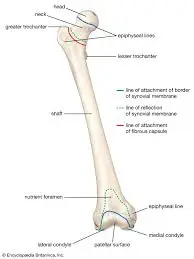
The femur has an extended shaft in the epicenter and two ends with rounded edges. It is the traditional cartoon bone shape: Two rounded bumps on either end of a cylinder. Despite being a single lengthy bone, your femur comprises several portions. These consist of:
The proximal portion of the femur:
The upper (proximal) end of your femur is where the hip joint attaches. The proximal end (aspect) comprises the following:
- Head.
- Neck.
- Greater trochanter.
- Lesser trochanter.
- Intertrochanteric line with crest.
Shaft of the femur:
The shaft is the long section of the femur that makes up your thigh and carries your weight. It leans slightly in the direction of your body’s center. Your femur’s shaft consists of the following:
- Linea aspera.
- Gluteal tuberosity.
- Pectineal line.
- Popliteal fossa.
The distal portion of the femur:
The upper aspect of your knee joint resides in the distal (base) region of your femur bone. It links your tibia (shin) and patella. This consists of the following:
- The medial and lateral condyles.
- Medial and lateral epicondyles.
- Intercondylar fossa.
Your medical professional will often rely on these sections and descriptions to indicate where you are feeling uncomfortable or having difficulty. Your doctor may use some of the following terms to describe the location of the bone break if you have a femur injury (a femur fracture).
Blood Supply:
- The femoral artery serves as the femur bone’s blood supply. The femoral artery branches off the obturator artery, which flows through the ligamentum teres femoris, to provide blood supply to the femoral head, which was but is not the primary source of development.
- The perforating branches of the deep femoral artery blood supply simultaneously the shaft and the distal region of the femur bone.
Muscles:
- The thigh muscles are separated into four sections.
- The anterior,
- The medial,
- The posterior and
- Gluteal regions.
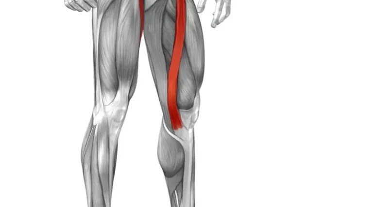
- The anterior compartment contains the femur. The hip flexor muscles and knee extensor muscles produce up the anterior part. The iliopsoas, sartorius, and pectineus muscles are collectively referred to as hip flexors.
- The medial compartment’s principal role is to adduct the legs.
- The most elementary muscle groups in the rare are the hip extensors and knee flexors.
- The outermost layer of muscular tissue is made up of the gluteus maximus, medius, and minimus. The superficial gluteal muscles’ principal functions are hip extension, abduction, and internal rotation.
- The layer that is most firmly buried is represented by the quadratus femoris, piriformis, obturator internus, and superior and inferior gemellus.
Articulations:
- The proximal femur’s femoral head as the ball the pelvic acetabulum as the socket, articulates therefore. Makes it easier to move the hips in the frontal (abduction and adduction), sagittal (flexion and extension), and horizontal (internal and external rotation) planes.
- The femur’s convex femoral condyles interact with the tibia’s condyles distally, forming the tibiofemoral joint. The two fundamental dimensions of motion of the tibiofemoral joint are internal and external rotation in the horizontal plane and knee flexion and extension in the sagittal plane.
- A patellofemoral joint happens when the patella communicates with the intercondylar and trochlear groove of the femur bone. When the knee is flexed and extended, the patella and femur’s articular surfaces slide together.
The function of the Femur
Your femur performs several vital functions, such as:
- Supporting your body weight while standing and moving.
- Maintaining your stability as you travel.
- Attaching the muscles, tendons, and ligaments of your knees and hips to the rest of your body.
- Adult femurs typically measure around 18 inches in length. It may withstand up to 30 times your body weight.
Affecting disorders and conditions of the Femur
Osteoporosis, patellofemoral pain syndrome, and fractures are among the many common conditions affecting the femur.
Fractures of the femur
In medical terms, breaking a bone is known as a bone fracture. Due to their extreme strength, femurs are typically only fractured by severe trauma, falls, or auto accidents. A fracture can be identified by symptoms such as those listed below:
- Pain
- Swelling
- Tenderness
- Being unable to move your leg as you typically do
- Discoloration or bruises
- a lump or malformation that is not typically present in your body
You should visit the emergency room quickly if you feel that you may have been damaged or have a fracture.
Osteoporosis
Bones weakened by osteoporosis are more prone to unexpected and abrupt fractures. When osteoporosis causes a bone to break, plenty of individuals are not aware that they have the condition. The symptoms are typically not noticeable straight away.
Osteoporosis is more likely to occur in women, those assigned female at birth, and persons over 50. Discuss with your healthcare provider the possibility of doing a bone density screening to detect osteoporosis before it results in a fracture.
Patellofemoral pain Syndrome
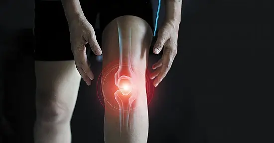
The condition known as patellofemoral pain syndrome (PFPS) is pain under and around your kneecap (patella). Runner’s knee or jumper’s knee are other names for it. Anything from acquiring new shoes to overusing your knees can result in PFPS. PFPS symptoms include:
- Pain in the knees when twisting, like in squatting or climbing stairs.
- Discomfort when bending your knees while sitting.
- When you stand up or go upstairs, you may hear crackling or popping sounds in your knee.
- Changes to your typical playing surface, sporting goods, or level of activity intensity exacerbate pain.
If you have recently developed knee pain, consult your healthcare physician.
Diagnosis for the Femur
A bone density test is the most popular procedure used to assess the condition of your femur. Sometimes, it is called a DXA or DEXA scan. A bone density test uses small amounts of X-rays to calculate how strong your bones are. It acts as a barometer for aging-related bone loss.
Your orthopedic surgeon or medical specialist could need imaging tests if you have had a femur fracture, including
- X-rays.
- MRI, or magnetic resonance imaging.
- CT scan.
Treatments for the Femur
Generally speaking, you don’t need to treat your femur in general. Treatment is required only if you have osteoporosis or have a fracture.
Treatment for femur fractures
The type of fracture you have and its etiology will determine how it is treated. To realign (set) your bone to its proper position and secure it in place so it can heal, you will likely require surgery in addition to some kind of immobilization, such as a cast or splint.
Treatment for osteoporosis
Vitamin and mineral supplements, medication, and exercise are all options to treat osteoporosis. Osteoporosis may typically be avoided with exercise and supplementation. With the help of your physician, a treatment strategy specific to your circumstances and bone health will be developed.
Maintaining the Health of Your Femur
You may maintain your bone (and general) health by adhering to a healthy diet and exercise regimen and scheduling routine checks with your healthcare professional. If osteoporosis runs in your family or you are over 50, ask your doctor for a bone density screening. Follow these general safety guidelines to reduce your risk of trauma:
- Wear a seatbelt at all times.
- When participating in any sport or activity, wear the proper protective equipment.
- Make sure there is nothing that could trip you or others in your house or place of work.
- At all times make use of the right equipment or tools when approaching something at home. Steer away from standing on tables, chairs, or workstations.
- Adhere to a diet and activity regimen that will support bone health.
- Carrying a cane or walker is recommended if you have difficulty walking or are more likely to fall.
Summary
Through your femur, you have a leg to stand on. The most extensive and most rigid bone in your body, it is also one of the most valuable and significant. Your bones will remain strong if you take any steps to enhance your general health.
FAQs
What is the femur bone?
Your thigh bone is called the femur. Your ability to move and stand is determined by it. Numerous vital muscles, tendons, ligaments, and components of your circulatory system are also supported by the femur.
What is the severity of a femur injury?
A smashed femur is a dangerous injury that has to be treated quickly. Physical therapy and surgery are the two main treatments for broken femurs. The healing process for a broken femur may take months. A car accident, a tumble, or a gunshot wound can all break your femur.
Is it possible to heal a femur without surgery?
The majority of patients who have a femur fracture require surgery, typically ORIF. Your shattered femur might not heal properly if you don’t get the procedure. Your bones can be repositioned correctly with ORIF. The likelihood that your bone will recover properly is greatly increased by doing this.
Is it possible to walk with a broken femur?
Normally, you can’t walk on it. To see the break, a doctor will take x-rays and maybe a CT scan. To relieve the pain and maintain the stability of your leg, they may place you in a knee brace.
What is the most effective way to treat a fractured femur?
Both surgical and non-operative methods can be used to treat femoral shaft fractures. In high-income nations, intramedullary nailing combined with surgical fixation is the gold standard of care. External fixation and plate osteosynthesis are further surgical methods.
References:
- Professional, C. C. M. (2024a, May 1). Femur. Cleveland Clinic. https://my.clevelandclinic.org/health/body/22503-femur
- The Healthline Editorial Team. (2018, January 20). Femur. Healthline. https://www.healthline.com/human-body-maps/femur#1
- Rohit, B. (2023, April 4). Femur Bone – Proximal, Distal, Shaft – Anatomy, Function. Samarpan Physiotherapy Clinic. https://samarpanphysioclinic.com/femur-bone/
- Vataliya, D. (2024, January 31). Femur Bone – Surprising Secrets of Your Thighbone. Mobile Physiotherapy Clinic. https://mobilephysiotherapyclinic.in/femur-bone/

One Comment