Anterior Talofibular Ligament (ATFL) Tear
What is an anterior talofibular ligament tear?
Anterior Talofibular Ligament tears (ATFLs) are among the most common ankle injuries, especially among athletes and people who participate in physical activities. These injuries can have a major impact on mobility and need to be properly understood, treated, and healed to heal. In this blog, we go over the recovery process for ATFL ligament tears, the timeframe for healing ATFL injuries, and the crucial safety measures to help with recovery.
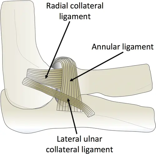
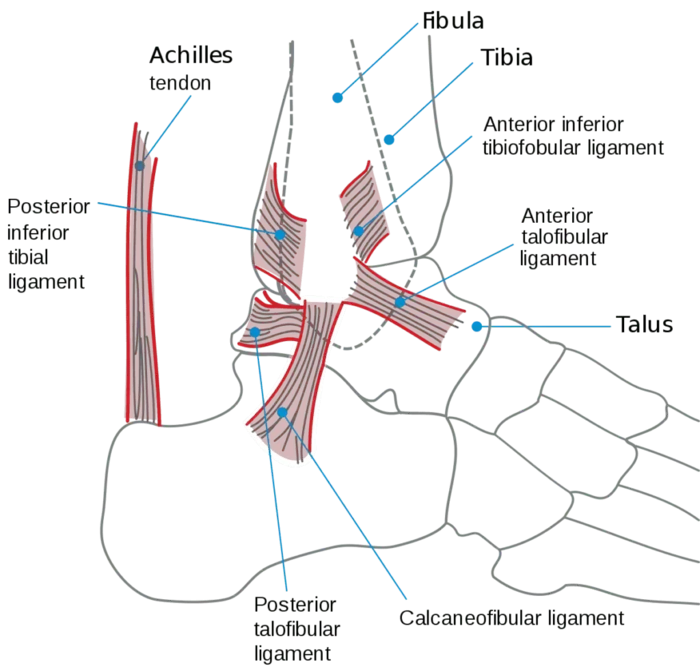
The lateral malleolus, neck, and talus are all connected by this ligament, which is located on the lateral part of the ankle. The primary function of the anterior talofibular ligament is to stop the foot from sliding forward. These injuries happen when the foot is flexed downward toward the ground and the ankle joint is inverted.
Clinical relevance Anatomy:
The most prevalent kind of acute sports injury is an ankle ligamentous sprain. According to reports, the most frequent injury among collegiate athletes in the US is a sprain of the ankle ligament.
Causes of anterior talofibular ligament tear:
- Injury and Trauma: These may be caused by severe pressure, twists, or unexpected ankle strikes. These movements overstretch or tear the ligament, forcing the ankle into an abnormal position.
- Sports-related Injuries: Sports like basketball, soccer, and volleyball that demand fast direction changes, leaps, and stops are common causes of ATFL injuries. These motions raise the possibility of ankle inversion, which might result in tears or sprains. These sports put a lot of strain on the ligament, which can lead to injury.
Symptoms of anterior talofibular ligament tear:
The anterior talofibular ligament can sustain a low ankle injury in three different degrees. Other typical signs of a tear in the anterior talofibular ligament include:
- Pain: An ATFL injury is frequently indicated by an abrupt flare-up of ankle pain. It can be severe and piercing, and it frequently gets worse when you move or change how much weight is supported on the injured ankle.
- Swelling: Ankle edema that occurs quickly is an indication of an ATFL injury because of the increased blood flow and fluid collecting at the injury site. The body uses this inflammatory reaction to prevent the injured area from more injury, even though it can be extremely uncomfortable, limit range of motion, and show the extent of ligament damage.
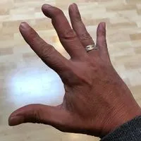
- Bruising: Blood pools beneath the skin as a result of tiny blood vessels bursting surrounding the injured ligament in an ATFL injury, causing bruises. Usually, this discoloration shows up a few hours to many days after the accident. An ATFL rupture or injury may be indicated by bruising around the ankle.
- Tenderness: An ATFL injury is indicated if your ankles feel more sensitive and painful than normal or if you touch or apply pressure to the ankle joint.
- Instability: An ATFL injury may also be indicated by unsteadiness or an inability to maintain weight appropriately following an ankle injury. This happens when there is insufficient ligament support, which might impair the stability of the ankle joint when moving.
- Limited Range of Motion: This limitation is frequently caused by joint pain, edema, and stiffness.
- Popping Sensation: An ATFL injury may provide a “popping sensation” after the first injury or during subsequent motions. This typically indicates ligament instability or structural displacement in the ankle joint, which can cause pain and edema.
Diagnosis:
To assess each ligament’s integrity, the lateral ankle ligaments undergo several particular procedures. A positive anterior drawer test indicates damage to both the calcaneofibular and anterior talofibular ligaments. Compared to patients with whole anterior talofibular ligament ruptures, individuals with partial tears may exhibit greater ankle joint stability. The extent of damage to each ligament and other soft tissue components of the ankle joint may be seen with this sophisticated imaging.
Assessment:
- Observation: Conduct a neurovascular evaluation and keep an eye out for any obvious anomalies, edema, or ecchymosis.
- Palpation: Check the ATFL and other lateral collateral ligaments for any sore spots. Examine the capillary refill, dorsal pedal pulse, and mild touch sensitivity. The medial malleolus, lateral malleolus, navicular, and base of the fifth metatarsal can all be included in a figure-8 assessment of edema using a tape measure.
- Range of Motion: Both the active and passive ranges of motion should be measured bilaterally using goniometry.
Special Tests:
Anterior Drawer Test: Hold the foot in a 20° plantar flexion position with one hand while using the other to bring the talus forward in the ankle mortise and stabilize the tibia and fibula. During the test, there can also be a noticeable clunk. Four to five days after injury, the anterior drawer test has been observed to have a significantly higher sensitivity due to acutely heightened pain and swelling.
Talar Tilt Test: Although the primary purpose of this test is to assess the calcaneofibular ligament’s (CFL) integrity, it can also provide important insights into the ATFL. A CFL injury is indicated by a positive test result, which is 5° to 10° of greater inversion relative to the non-injured ankle.
Classification of anterior talofibular ligament tear:
- Grade 1: A little ATFL tear.
- Grade 2: The ATFL has a large tear. The anterior joint capsule, ATFL, and adjacent soft tissue components are torn, resulting in bruising, localized swelling, a painful gait or incapacity to walk, and a wider area of point soreness over the lateral part of the ankle.
- Grade 3: The ATFL completely ruptures, maybe involving the CFL. The symptoms include pain on the medial and lateral sides of the ankle joint, incapacity to walk, and widespread swelling that completely covers the Achilles tendon’s boundaries.
Treatment of anterior talofibular ligament tear:
Medical Treatment:
Patients with anterior talofibular ligament tears typically only require conservative therapy for these kinds of injuries. Being assessed by a foot and ankle specialist as soon as possible after the injury is crucial because early management produces the best results.
With these injuries, regular rest, ice, elevation, and compression can also help reduce pain and swelling. It has also been demonstrated that a muscular strengthening-focused physical rehabilitation program aids patients in getting back to their regular physical activities.
Acute Inflammatory Phase:
Grade 1 and 2 Sprains:
- Rest or Adjusted Activity: As much weight as tolerated
- Active Range of Motion (ROM): To reduce inflammation and improve circulation, patients should be taught to do ankle pumps (10 to 20 per hour) within a pain-free range.
- To promote early ligament healing, soft tissue treatments such as Active Release treatments, Graston Technique, muscular energy techniques, and transverse friction massage can be given directly to the ligament and surrounding soft tissue structures.
Grade 3 Sprains:
Suspicion of a grade 3 sprain must be justified if the patient is unable to support their own weight and exhibits severe ankle pain and edema at the time of initial assessment. After that, the patient should be told to follow the RICE procedure (Rest, Ice, Compression, Elevation) until the MRI is ready.
Reparative Phase:
- Reduce inflammation
- Strength Training
- Maintain cardiovascular fitness
- Proprioceptive rehabilitation
- Stabilization
Remodeling Phase:
- Advanced strength training
- Agility Training
- Multi-directional sports-specific proprioceptive training
Surgical Treatment:
Surgery may be necessary to stabilize the ankle joint in patients who do not respond to conservative treatment or in certain cases where the ligament has completely ruptured. The ATFL ligament injury may require surgery to be repaired. The ligament’s remaining healthy tissue is sutured back together or reattached to the bone after the damaged sections have been cleansed. The type of tear determines the precise incision and method.
Precautions for Anterior Talofibular Ligament Tear Recovery:
Certain safety measures are essential for promoting a speedy recovery from ATFL tears and lowering the chance of complications or re-injury:
- Weight Management: During the initial healing phase, avoid placing too much weight or pressure on the injured ATFL ankle; crutches or a supportive walking aid may be required.
- Physical Therapy: Participate in a structured physical therapy program that emphasizes ankle strengthening, range of motion exercises, and proprioception training to restore stability and function.
- Immobilization: Adhere to the recommended period of immobilization, whether using a brace, splint, or cast, to allow the ATFL ligament tear to heal without undue stress.
- Gradual Return to Activity: Under the supervision of a medical specialist, gradually resume weight-bearing exercises and sports-specific drills. Resuming demanding activities too soon can compromise recovery and result in repeated injuries.
- Appropriate Footwear: When participating in sports or other physical activity, wear supportive, well-fitting shoes that can offer sufficient ankle support and cushioning.
- Avoid away of risky movements: Activities that require abrupt direction changes, jumping, or uneven surfaces should be avoided as they may cause strain on the repaired ligament.
- Regular Follow-ups: Keep track of your progress, discuss any issues, and modify the treatment plan as necessary by attending your doctor’s appointments on time.
Prevention of Anterior Talofibular Ligament (ATFL) Tear:
Maintaining ankle strength, flexibility, and stability is crucial for preventing an anterior talofibular ligament (ATFL) tear, particularly in athletes and those who engage in other activities that put strain on the ankle. By strengthening neuromuscular control, regular workouts that improve balance and proprioception can dramatically lower the incidence of ankle sprains.
Ankle braces or taping can be used to give extra support and stop re-injury for people who have previously suffered ankle injuries. The risk of an ATFL tear can also be reduced by warming up before exercise, avoiding uneven surfaces, and progressively increasing the intensity of physical activity.
Complications
If an anterior talofibular ligament (ATFL) tear is not appropriately treated or if rehabilitation is insufficient, complications may result. Chronic ankle instability, when the ankle feels weak or gives way, especially when exercising, is one of the most frequent side effects. Recurrent ankle sprains, decreased sports performance, and trouble walking on uneven surfaces might result from this.
Post-traumatic osteoarthritis in the ankle joint may develop over time as a result of instability and recurrent trauma. Persistent pain, edema, and stiffness are other possible side effects. Weakened proprioception can also lead to restricted range of motion and poor balance. To reduce these dangers and guarantee a full recovery, an organized rehabilitation program and early diagnosis are crucial.
Prognosis:
Anterior Talofibular Ligament (ATFL) tears often have a good prognosis, especially when treated appropriately and promptly. Most people can resume their regular activities in a few weeks after mild to moderate tears (Grades I and II) are treated conservatively with rest, ice, compression, elevation (RICE), and physical therapy.
A longer recovery time may be required for severe tears (Grade III), and surgery may be required in cases of persistent instability or repeated sprains. Most patients recover completely and regain their pre-injury function with appropriate therapy that emphasizes strength, stability, and proprioception; nevertheless, certain individuals may be at risk for chronic ankle instability if their condition is not appropriately treated.
Conclusion:
In conclusion, several variables, including the extent of the tear, the course of therapy, and adherence to safety measures, affect how long it takes to recover from an ATFL injury. More serious injuries may necessitate months of therapy and potentially surgery, whereas partial ligament tears may mend with conservative methods in a matter of weeks.
To get the best results and avoid more ankle issues, it is essential to take care, heed medical instructions, and exercise patience while recovering.
FAQs
Is it possible to walk on an ATFL sprain?
While a grade 2 or grade 3 sprain usually causes pain and instability during any weight-bearing exercise, a grade 1 sprain may nevertheless permit a patient to walk without pain or instability. After an injury, rest is recommended for at least 24 hours, but exercise can begin shortly after.
How can an ATFL tear be fixed?
The ATFL and CFL ankle ligaments may be removed by your surgeon from their attachment points on your fibula. They might shorten these ligaments. If more repairs are required, your surgeon may perform them.
Why do ATFL injuries occur most frequently?
Of the three ligaments, the ATFL is the weakest and shortest, with a limited ability to sustain maximum strain until collapse. Consequently, up to 85% of all ankle sprains include the ATFL, making it the most often injured ligament during an inverted ankle sprain.
How can an ATFL tear be repaired?
The ATFL ligament injury may require surgery to be repaired. The ligament’s remaining healthy tissue is sutured back together or reattached to the bone after the damaged sections have been cleansed. The type of tear determines the precise incision and method.
Can someone who has a torn ATFL walk?
The extent of ligament damage will determine whether the patient has mild, moderate, or diffuse swelling on the outside of the ankle. Patients with minor injuries may be able to bear their entire weight, but those with severe ankle ligament damage may not be able to put their foot down at all.
Is surgery necessary for an ATFL tear?
However, surgical ligament rebuilding will be recommended if this is ineffective and you continue to experience significant pain and joint instability.
What is the ATFL operation success rate?
Many patients regain ankle stability and resume their prior activity levels following ATFL reconstruction procedures, which typically have high success rates.
Is surgery necessary to repair a torn ATFL?
No, surgery is not required for every torn ATFL injury. If conservative measures are ineffective in restoring function and stability, or if the ATFL ligament is completely torn, surgery may be required.
References
- Drhaytmanek. (2025, January 6). Anterior Talofibular Ligament (ATFL) Tear. Sport Foot & Ankle. https://sportsfootankle.com/anterior-talofibular-ligament-atfl-tear/
- Chen, R., Wang, Q., Li, M., Su, X., Wang, D., Liu, X., & Li, Z. (2023). Progress in diagnosis and treatment of acute injury to the anterior talofibular ligament. World Journal of Clinical Cases, 11(15), 3395–3407. https://doi.org/10.12998/wjcc.v11.i15.3395
- Sheikh, Y., & Weerakkody, Y. (2013). Anterior talofibular ligament injury. Radiopaedia.org. https://doi.org/10.53347/rid-24471
- Prasad, N., & Prasad, N. (2025, April 8). Anterior talofibular ligament (ATFL) injury treatment. The Cruciates – The Cruciates Centre of Excellence for all kinds of Sports Injuries. https://thecruciates.com/understanding-atfl-injuries-causes-types-healing-time-and-precautions/
- Lai, M. W. W., & Sit, R. W. S. (2018, March 1). Healing of Complete Tear of the Anterior Talofibular Ligament and Early Ankle Stabilization after Autologous Platelet Rich Plasma: a Case Report and Literature Review. https://pmc.ncbi.nlm.nih.gov/articles/PMC5867359/