Atlantoaxial Joint
The atlantoaxial joint is in the upper neck region between the first and second cervical vertebrae or the atlas and axis bones. The joint is essential.
The synovial joint known as the atlantoaxial joint is categorized as a uniaxial pivot joint. It is classified as a pivot joint since it comprises a centrally located bony component that pivots around the craniovertebral ligaments and only permits rotation.
Introduction
The atlantoaxial joint (AAJ), which connects the first cervical vertebra (C1) to the second cervical vertebra (C2), is the most flexible part of the spine. Two lateral plane joints and one median pivot joint make up this joint’s three synovial joints. Approximately 50% of cervical rotation is produced by this intricate structure, which provides stability while permitting significant flexibility.
It is susceptible to some diseases, such as instability, degenerative changes, and dislocation, due to its distinct structure.
Rotation is the main motion of the atlantoaxial joint complex. The head and the atlas spin around the axis of rotation. With this movement, we can turn our head to see either left or right. We can also shake our heads in the well-known “no” pattern with this motion.
Structure and Function
With several different structural features that contribute to its vital role in cervical stability and mobilization, the atlantoaxial joint is a special kind of joint. Two lateral atlantoaxial joints and one medial joint make up its three separate synovial joints. The anterior and posterior ligamentous structures, as well as the dens of the C1 vertebrae, create the structure of the median. These are, respectively, the transverse ligament and the atlas’ osteoligamentous rings. Classified as gliding joints are the two bilateral atlantoaxial joints seen laterally.
The atlantoaxial joint is composed of the axis (C2) and the atlas (C1), the top portions of the cervical spine. These two vertebrae are distinct from the other vertebrae in the cervical spine because of their particular anatomical structure, which is found near the craniovertebral junction, where the skull and cervical spine converge. This joint differs from the others in the cervical spine because it does not have an intervertebral disk.
The atlantoaxial joint’s multifunctionality, which enables the head to rotate and turn to the left or right, is essential in real-world applications. The kinematic capabilities of the head and neck would not be able to support the over 600 movements per hour that humans perform without the atlantoaxial joint. This cervical spine joint has a variety of functions. The head is generally stabilized and the weight is supported in a neutral position. Additionally, this cervical unit helped shield the spinal cord from outside pressure.
Additionally, it permits the vertebral artery to carry blood to the brain. Along with protecting and stabilizing the head, neck, and internal tissues, the atlantoaxial joint also helps the neck muscles and head-neck movements. A capsule of articular cartilage surrounds the atlantoaxial joint as well, serving to connect the posterior surface of the axis to the atlas margins at the lateral masses.
Atlas (C1)
The atlas is a round structure that articulates with the cranium above it through lateral masses or zygapophyseal joints. Through its bilateral condyles, the atlas articulates with the axis below. Another feature that sets the atlas apart from the other cervical vertebrae is the absence of a spinous process or vertebral body. The transverse ligament, which serves as a groove for the vertebral artery, is also found in the atlas between the anterior and posterior arches, two superior articular surfaces.
Axis (C2)
This vertebra is known as the C2 vertebra and is distinguished by the dens or odontoid process. This structure extends superiorly from the front section of the vertebrae and articulates with the axis above. The bilateral transverse foramina, bifid spinous process, and superior articular facets are further axis traits.
Embryology
Blastocyst development is the result of several cellular divisions that take place throughout pregnancy during the first two weeks after conception. The process of gastrulation, which involves the blastocyte forming into a stratified structure made up of the ectoderm, mesoderm, and endoderm, occurs near the end of the second week. Mesodermal structures termed somites start to migrate into their mature, functional form as vertebrae during the establishment of the cervical spine and atlantoaxial joint.
The notochord involutes and somites develop into their mature forms at primary ossification centers, which are vertebrae. The following processes occur during normal cervical spine development in embryogenesis: gastrulation and somatic mesodermal creation, mesoderm condensation into somites, dermomyotome and sclerotome reorganization, somite segmentation, chondrification, and ossification of the vertebrae. One ossification center for the vertebral body, two for each of the neural arches, and one for the dens are all located along the axis.
Since the junction between the dens and the body of the axis does not fully fuse until around age six, these dens ossification foci are especially significant because they can be mistaken for fractures in juvenile patients. The odontoid process and the axis body are known to unite by the ages of 4 and 6 and to fully develop by the age of 25.
Lateral atlantoaxial joints
Articular surfaces
The inferior articular surface of the lateral mass of the atlas (C1) and the superior articular surface of the lateral mass of axis (C2) articulate to form the left and right lateral atlantoaxial joints. Hyaline cartilage covers the articular surfaces of these joints because they are synovial.
Even though they are categorized as planar-type joints, the lateral atlantoaxial joints have somewhat more complicated articular surfaces. Their long axis runs from anteromedial to posterolateral at an oblique angle, and both the axis and atlas have oval articular surfaces.
Additionally, the surfaces have a slight inferior tilt, with the anterior aspect being superior to the posterior. Finally, flexible meniscoids cover the anterior and posterior joint edges that are not congruent, and the articular surfaces are convex in the sagittal plane.
Joint capsule
The loose fibrous capsules that enclose the lateral atlantoaxial joints are coated with a synovial membrane. The lateral masses of the axis and atlas are enclosed by the joint capsule, which is attached to the lateral articular facets of the joints. An auxiliary atlantoaxial ligament reinforces each side of the joint capsule.
Ligaments
The posterior aspect of the lateral atlantoaxial joints is supported by the supplementary atlantoaxial ligament. It originates from the tectorial membrane’s deep laminae and extends laterally. On the posterior face of the atlas’ lateral mass, the ligament blends with the fibers of the posterior capsules and attaches superiorly close to its transverse ligament. From there, it travels medially and obliquely downward to join the rear of the axis body close to the dens’ base.
Although its precise involvement in rotational stability at the craniocervical junction is unknown, the auxiliary atlantoaxial ligament is believed to be involved. It should be mentioned that others consider these fibers to be a part of the tectorial membrane rather than a distinct ligament.
Median atlantoaxial joint
Articular surfaces
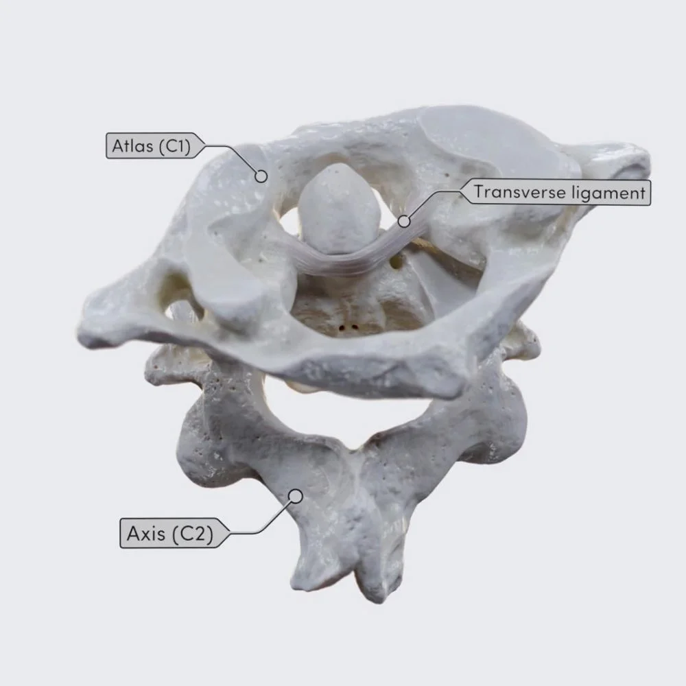
A pivot-type synovial joint is the median atlantoaxial joint. In general, the odontoid process, or dens of the axis, forms the joint. It is encircled by an osteoligamentous ring made up of the transverse ligament of the atlas posteriorly and the anterior arch of the atlas anteriorly.
There are two sets of articulations within this osteoligamentous ring, each with its synovial cavity. The inner surface of the anterior arch of the atlas (cervical vertebra 1) and the anterior articular facet of the dens of the axis (cervical vertebra 2) articulate with one another. The transverse ligament of Atlas’s front surface and the posterior articular facet of the dens of axis form the second articulation.
The dens’s anterior facet is rectangular and convex in both the transverse and vertical planes. It fits against the atlas’s front arch’s appropriately concave surface. Concave vertically and convex transversely are the dens’ rear articular faces. The transverse ligament’s fibrocartilaginous surface is where it rests.
Hyaline cartilage covers every articulating surface of the median atlantoaxial joint.
Joint capsule
There are two synovial cavities in the median atlantoaxial joint, one on either side of the dens of the axis. Between the dens and the transverse ligament, the larger of the two is the posterior synovial cavity. The synovial membrane lines a thin joint capsule that encloses each synovial cavity. A significant range of motion is possible within the joint due to the relative looseness of this joint capsule, particularly in the superior section.
Ligaments
Many ligaments hold the median atlantoaxial joint in place. The atlas and axes are connected by the primary ligaments of the joint, which are referred to as the cruciform ligament complex.
Three ligaments—two longitudinal and one horizontal—combine to form the cruciform ligament, which gets its name from the way they resemble a cross. The following are the three bands that make up the cruciform ligament:
- Atlas’s transverse ligament is a robust, wide ligament that connects to tubercles on their medial faces and runs transversely between the atlas’ lateral masses. Also known as the transverse atlantal ligament, it broadens in its central region, where a layer of articular cartilage covers it anteriorly, and arches behind the dens. Through its posterior articulation, this ligament contributes to the development of the osteoligamentous ring surrounding the dens of the axis. The transverse ligament, which acts to prevent the atlas from translating forward to the axis, is the primary stabilizing factor of the dens of the axis.
- The superior longitudinal band of the cruciform ligament extends upward to insert at the basilar portion of the occipital bone from the superior margin of the median portion of the transverse ligament of the atlas. The tectorial membrane and the dens’ apical ligament are where the attachment is located.
- The cruciform ligament’s inferior longitudinal band descends to attach on the posterior aspect of the body of the axis after emerging from the inferior edge of the transverse ligament of the atlas’ median portion.
The median atlantoaxial joint has multiple accessory ligaments in addition to its primary ligaments that unite the occipital bone and the axis (C2).
- The cervical vertebral column’s tectorial membrane is the superior extension of the posterior longitudinal ligament. This robust, wide band begins on the back side of the axis’s body and rises to insert on the foramen magnum’s anterior border. Attached close to the hypoglossal canals, it blends in with the meninges or spinal dura mater. The posterior surface of the dens is covered by the tectorial membrane. After the cruciform and alar ligaments, it is located.
- Alar ligaments: these two ligaments join to the medial portions of the occipital condyles after an oblique, superolateral path from the posterolateral borders of the apex of the dens of the axis (C2). The short but robust alar ligaments restrain excessive motion in the atlantoaxial joint.
- Apical ligament of dens: attaches to the anterior edge of the foramen magnum by branching out superiorly from the apex of dens. The cruciform ligament’s superior longitudinal band is in front of the apical ligament, which inserts between the two alar ligaments. Also named the apical oral ligament, this reflects the vestigial cranial extension of the notochord. Its biomechanical importance is debated.
- The anterior atlantoaxial membrane, which extends from the inferior border of the atlas to the inferior portion of the axis, is a continuation of the anterior longitudinal ligament. Continues as the anterior atlanto-occipital membrane to the occiput.
- A superior continuation of the ligamentum flavum, the posterior atlantoaxial membrane spans the space between the atlas and axis, running from the upper margins of the axis’s laminae to the inferior border of the atlas’s posterior arch. The posterior atlanto-occipital membrane extends to the occiput.
Innervation
The ventral major ramus of the second cervical spinal nerve innervates the atlantoaxial joint by branches.
Blood supply
Branches of the deep cervical, occipital, and vertebral arteries anastomose deliver arterial blood to the atlantoaxial joint.
Movements
One of the most flexible joints in the spine is the atlantoaxial joint. It enables us to shake our heads in the well-known “no” pattern or swivel our heads to look left and right. All three of the atlantoaxial joints—the medial and two lateral—act together to produce axial rotation, which is the movement’s result.
The right lateral mass of the axis translates anteriorly on the articular facet of the axis in the case of the left rotation. The left lateral mass of the axis exhibits reciprocal posterior displacement at the same moment. These motions take place at the lateral atlantoaxial joint of the plane type. Concurrently, the dens, which are contained inside its osteoligamentous ring, are centered by the atlas. The head turns leftward as a result of the head and atlas (C1) moving as a single unit over the axis (C2).
Rotation to the right does the opposite. At the atlantoaxial joint, the rotational range of motion is 40° (range 39-49°). A modest “screwing down” of the atlas on the axis occurs during rotation because of the convex, obliquely oriented articular surfaces, where the atlas drops vertically by about 1 millimeter. The alar ligaments primarily restrict rotational mobility.
There is also limited flexion, extension, and lateral flexion possible at the atlantoaxial joint. The front arch of Atlas rolls and slides somewhat on the dens of the axis to induce flexion and extension. The atlas glides anteroinferiorly on the axis during flexion, and there is also a small anterior translation. A slit backward tilting is made possible by the atlas sliding superoposteriorly in extension. The weak and loose joint capsule and the moderately flexible transverse ligament, which bends upward during extension and downward during flexion, allow for these movements.
According to reports, the range of flexion-extension motion is 11–21°. With a reciprocal displacement on the other side, lateral flexion is created when the inferior articular facet slides along the convex oval facet on one side. As a result of the inferior inclination of the joint surfaces, the atlas experiences a slight lateral tilt, which causes lateral bending on the axis. It has been demonstrated that rotation and contralateral lateral flexion are related. Atlantoaxial joint lateral flexion has a reported range of motion of only 5 to 10°.
The entire atlantoaxial joint is in an open-pack position when the head and neck are semiflexed, and it is in a closed-packed position when the head is fully extended. Equally limited lateral flexion, extension, and rotation are described by the atlantoaxial joint’s capsular pattern. Even though we can take into account the segmental motion at the C1-C2 joint, craniovertebral movements are not isolated. Instead, the upper cervical spine as a whole works as a single joint complex.
Muscles acting on the atlantoaxial joint
The rectus capitis posterior major and ipsilateral obliquus capitis inferior are the two main suboccipital muscles that generate rotation in the atlantoaxial joint. In addition, the contralateral sternocleidomastoid muscle and the ipsilateral splenius capitis muscle are engaged.
The suboccipital muscles aid in posture maintenance and rotation, as well as proprioception. The obliquus capitis inferior, which attaches to the atlas’ transverse process, also stabilizes the atlantoaxial joint.
Clinical significance
A fracture or injury to the atlantoaxial joint can result in major issues due to its proximity to the brain stem and its significant instability. Typical trauma-related disorders consist of (but are not restricted to):
The dens can push into the brainstem and cause death if there is a large depression in the skull. The dens itself can break as a result of ossification or trauma.
Transverse ligament: If the atlas’s transverse ligament is damaged or diseased, the dens become unanchored and may glide up the cervical spine, paralyzing the affected area. Death may ensue if it gets to the medulla. Alar ligaments: The weaker alar ligaments can be stretched by stress or trauma, increasing the range of motion by about 30%.
Genetic characteristics may occasionally cause ossification of the posterior atlantooccipital membrane, transforming the groove into a foramen.
Arthritis
The atlantoaxial joint is susceptible to osteoarthritis. This includes the traditional pathophysiology, which includes bone thickening with limited joint space, radiographic osteophytes, and loss of articular cartilage. Conservative treatment, which includes analgesics, is typically successful. In severe cases, surgery may be used, and the results may be favorable.
Abnormal widening
The transverse atlantal ligament is injured when the atlantoaxial joint widens, as assessed by the distance between the front of the odontoid process and the posterior surface of the anterior arch of the atlas. Although a maximum of 3 mm for men and 2.5 mm for women is occasionally acceptable, the atlanto-dental distance is typically less than 2 mm.
FAQs
How does one define the atlantoaxial joint?
The synovial joint known as the atlantoaxial joint is categorized as a uniaxial pivot joint. This joint is situated between the first and second cervical vertebrae, also referred to as the axis and atlas, respectively, in the upper portion of the neck.
What joint does C1 have with C2?
The atlantoaxial joint (AAJ), which connects the first cervical vertebra (C1) to the second cervical vertebra (C2), is the most flexible part of the spine. This joint is made up of two lateral plane joints, one median pivot joint, and three synovial joints.
Why is C1 referred to as Atlas?
The most widely accepted reason for the name Atlas for the first cervical vertebra is because the vertebra supports the globe of the skull in the same manner that Atlas supports the globe of the sky. But the Titan was being punished, which is a significant aspect of the Atlas tale.
What does an atlas serve as?
While allowing for significant motion in flexion, extension, rotation, and lateral bending, the atlas and axis support the head on the lower cervical spine. Additionally, the cervical cord and vertebral arteries pass through the first two vertebrae.
What are the “yes” and “no” joints?
They are made up of two sets of joints: the atlantooccipital joints, which are referred to as “yes” or nodding movement joints, and the atlantoaxial joints, which are referred to as “no” or rotating movement joints, which are positioned between the atlas (C1 vertebra) and the axis (C2 vertebra).
Which neck joint is the highest?
These are, respectively, the transverse ligament and the atlas’ osteoligamentous rings. Classified as gliding joints are the two bilateral atlantoaxial joints seen laterally. In the cervical spine, the atlas (C1) and the axis (C2) are the highest segments that comprise the atlantoaxial joint.
For what reason is Atlas unique?
According to legend, Atlas was an expert in astronomy, mathematics, and philosophy. He was credited with creating the first celestial sphere in antiquity. According to some accounts, he even created astronomy itself.
What is an atlantoaxial joint?
single-axis pivot joint
The synovial joint known as the atlantoaxial joint is categorized as a uniaxial pivot joint. This joint is situated between the first and second cervical vertebrae, also referred to as the axis and atlas, respectively, in the upper portion of the neck.
What is the connection between AA and OA?
The topmost joints in the spine, located directly below the head, are called occipito-atlantus joints (OA) and atlanto-axial joints (AA). Damage to the cartilage, trauma, osteoarthritis, rheumatoid arthritis, and other conditions can cause arthritis discomfort in either joint.
What is the atlantoaxial joint’s issue?
Since the atlantoaxial joint is the spine’s most movable articulation, instability and dislocation are common. The possibility of compression of vascular and neurological systems makes atlantoaxial rotatory dislocation, which can happen in the context of both traumatic and nontraumatic occurrences, extremely dangerous.
References
- Forbes, J., & Das, J. M. (2023b, August 28). Anatomy, head and neck: Atlantoaxial joint. StatPearls – NCBI Bookshelf. https://www.ncbi.nlm.nih.gov/books/NBK563271/
- Atlantoaxial joint. (2023, October 30). Kenhub. https://www.kenhub.com/en/library/anatomy/atlantoaxial-joint
- Wikipedia contributors. (2024c, August 23). Atlanto-axial joint. Wikipedia. https://en.wikipedia.org/wiki/Atlanto-axial_joint