Colle’s Fracture
What is a Colle’s Fracture?
The Colles fracture, named for Abraham Colles, who initially reported a distal radius fracture in 1814 at the Royal College of Surgeons in Dublin, is one of the most frequent fractures seen in orthopedic practice, accounting for 17.5% (or one-sixth) of all adult fractures that present to the emergency room. A distal radius fracture with dorsal comminution, dorsal angulation, dorsal displacement, radial shortening, and an associated ulnar styloid fracture is referred to as a Colles fracture.
Distal fractures with dorsal angulation are frequently referred to by their eponym, Colles fractures. Many times, falling on an outstretched hand with the wrist in dorsiflexion strains the volar aspect of the wrist, causing the fracture to expand dorsally. This is how distal radius fractures occur.
Related Anatomy
The proximal side of the wrist joint is formed by the distal radius. There, the radius forms an articulated joint for flexion and extension with the proximal row of carpal bones and a joint for pronation and supination with the distal ulna.
A meniscus-like structure called the triangular fibrocartilage discus (TFC), to which the distal ulna is attached, is prone to tearing in the event of a wrist fracture.
The brachioradialis inserts into a styloid projection on the lateral side of the radius, which is also the source of the radial collateral ligament of the wrist.
The cortical thickness of the bone is smaller than the proximal and distal bone at the distal metaphysis of the radius, and the proportion of cancellous bone increases. Thus, the radius’s distal metaphysis is a rather weak location. As a result, fractures become more frequent, especially in people with decreased bone mineral density.
Distal radius extra-articular fracture with low energy may be connected to scapholunate dissociation, ulnar styloid fracture, and TFCC (triangular fibrocartilage complex) tear.
For additional details regarding the anatomy and evaluation of the wrist.
Causes of Colles Fracture:
The common cause of Colles fractures is trauma. This usually relates to sports-related injuries or auto accidents in younger individuals. Falls are the most frequent cause of this in the senior population. When a traumatic force impacts an outstretched hand, the colles fracture can cause dorsal comminution when it is broken and shifted dorsally.
The wrist is used to dorsally transmit the force. Due to this process, a well-known wrist deformity called the “dinner fork deformity” is produced, along with pain, swelling, and a decrease in range of motion.
A colon fracture can be related to many additional conditions, including triangle fibrocartilage injury (TFCC), lunotriquetral ligament, scapholunate, and distal radioulnar joint damage (DRUJ). A concomitant radial styloid fracture would be indicative of a high-energy cause of damage.
Osteoporosis is a common risk factor for wrist fractures. Because osteoporosis affects bones, less force is needed to break them. Sometimes a fractured wrist is the first sign of bone degeneration. A forceful wrist injury is the cause of a Colles wrist fracture.
This might occur because of:
- Distal radioulnar joint injury (DRUJ), triangular fibrocartilage injury (TFCC), scapholunate, and lunotriquetral ligament injuries are only a few of the injuries that can be linked to distal radius fracture. A concomitant fracture of the radial styloid would suggest a high-energy mechanism of injury.
- Accident with a vehicle
- contact Sports
- The most common cause of falling when riding, snowboarding, or engaging in other activities is tripping over an extended arm.
Pathophysiology:
Trauma is the most common cause of Colles fractures. In younger patients, this typically has to do with sports-related injuries or vehicle accidents. Falls are the most frequent cause of this in the senior population. The distal radius is fractured, shifted dorsally, and frequently results in dorsal comminution when a traumatic force strikes an outstretched hand. The force passes dorsally through the wrist.
This process results in the well-known wrist deformity characterized as the “dinner fork,” which is accompanied by discomfort, edema, and a decrease in range of motion
Distal radioulnar joint injury (DRUJ), triangular fibrocartilage injury (TFCC), scapholunate, and lunotriquetral ligament injuries are only a few of the injuries that can be linked to distal radius fracture. A concomitant fracture of the radial styloid would suggest a high-energy mechanism of injury.
Risk Factors of Colles’s Fracture:
Colles fractures are the most common type of distal radial fracture and affect all adult age groups and populations. These are highly prevalent in people with osteoporosis. As a result, older women are the most frequently observed, especially when they fall and have an outstretched hand.
A male patient who is older and has a Colles fracture should also be evaluated for osteoporosis because the two conditions have a strong connection and osteoporosis enhances the patient’s risk of hip fracture.
Because of growing awareness of osteoporosis, these wounds are now referred to as fragility fractures, indicating that an osteoporosis workup should be a frequent part of treatment. The longer people live, the more often this type of fracture occurs.
These fractures are more common in the elderly and in women, who are more likely to have osteoporosis due to aging-related osteoporosis. Osteoporosis is a risk factor for fragility fractures in the future, especially in women over 50. A distal radius fracture in a woman over 50 is ideal to detect in a DEXA scan to assess the bone’s integrity.
Younger people’s bones are stronger, thus it takes more force to fracture bones. In younger patients, high-impact injuries or falls—from contact sports, skiing, horseback riding, or motorcycle accidents, or falls from a high usually result in a clavicle fracture.
Types of Colles Fracture:
Based on the location and type of bone break, fractures will be classified. This helps determine what would be the best for you to do.
Here are a few forms of fractures:-
- Open fracture:- Is there evidence that the bone penetrated your skin
- A comminuted fracture:- occurred when there were more than two shattered fragments of bone.
- Intra-articular fracture:- if the fractured bone injured the wrist joint
- Extra-articular fracture: if your joint is unharmed
Classification:
| Type | Fracture |
| 1 | Extra-articular radial fracture. |
| 2 | Extra-articular radial fracture with an ulnar fracture. |
| 3 | Intra-articular fracture of the radio-carpal joint without an ulnar fracture. |
| 4 | Intra-articular fracture of the radius with an ulnar fracture. |
| 5 | Fracture of the radio-ulnar joint. |
| 6 | Fracture into the radio-ulnar joint with an ulnar fracture. |
| 7 | Intra-articular fracture involving radio-carpal and radio-ulnar joints. |
| 8 | An intra-articular fracture involving radio-carpal and radio-ulnar joints with ulnar fractures. |
Symptoms of Colles Fracture:
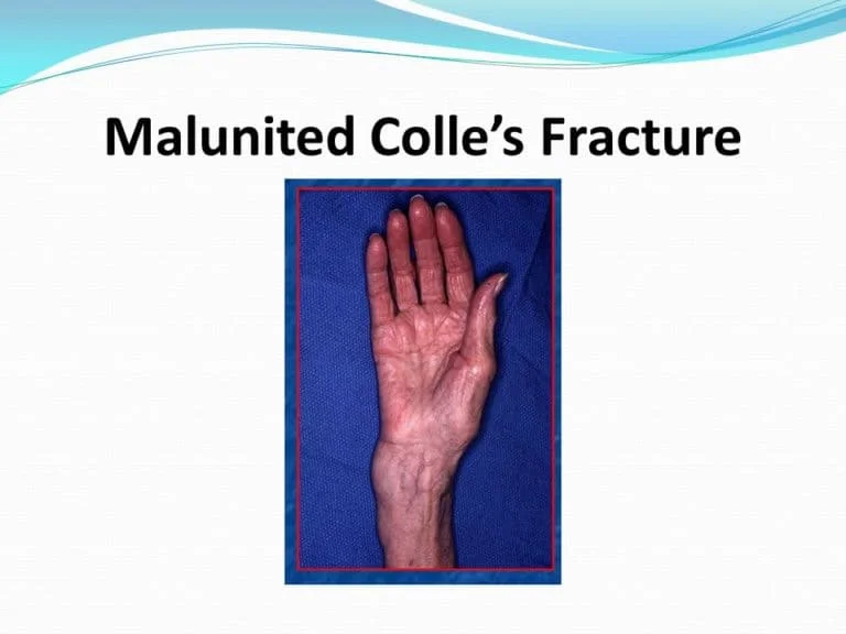
Clinically, colle”s fractures usually manifest as a fork-like deformity. A distal radius fracture causes a posterior displacement of the distal component, which brings the forearm posteriorly clinically, collapses fractured, and somewhat closer to the wrist.
In the typical forward arch of the hand, the patient’s hand and forearm resemble a dinner fork.
- Dinner-Fork deformity
- fall on an extended hand in history
- Dorsal wrist pain and wrist swelling Distal radius angulation elevation
- unable to grasp the thing
Common Signs of Colles Fractures:
You may develop a Colle’s fracture if you break your wrist or if you fall on the hand or wrist. Typical indications of a Colle’s fracture or wrist fracture include:-
- Pain
- swelling in the arm, wrist, or hand.
- bruising
- loss of wrist mobility
- An area that is visible on the back of your forearm, near your wrist.
- Tenderness
- wrist deformity
Diagnosis:
A thorough medical history that includes the mechanism of damage raises the possibility of a Colle’s fracture. Most typically, the diagnosis is based solely on the interpretation of the lateral and posteroanterior views.
The following characteristics relate to the classic Colle’s fracture:-
- 2.5 cm (0.98 inches) proximal to the radio-carpal joint dorsal displacement, dorsal angulation, and radial tilt, transverse fracture of the radius
Additional features that may be seen on plain radiographs include:
- Radial shortening
- ulnar inclination loss
- wrist radial angulation
- Crushing at the point of fracture
more than 60% of patients have an associated ulnar styloid process fracture.
Physical Examination:
Colle fractures require thorough physical examinations of both the damaged and unaffected extremities to be diagnosed and treated. There will be visible discomfort and wrist pain throughout the examination. Occasionally, an individual dorsiflexion deformity also referred to as a “dinner fork” deformity is seen when the fracture is displaced. There might also be evident bruises and swelling, and a thorough skin examination is necessary to determine if an open fracture is possible.
Even though the injury might have less range of motion, the doctor should however assess it if at all possible. Evaluating the neurovascular status of the extremity distal to the injury is essential, and it includes a careful assessment of the motor function, sensation, and pulses of the affected extremity. Joints above and below the injury must always be assessed to see if there are any connected injuries.
Investigation of Colle’s Fracture:
X-rays:
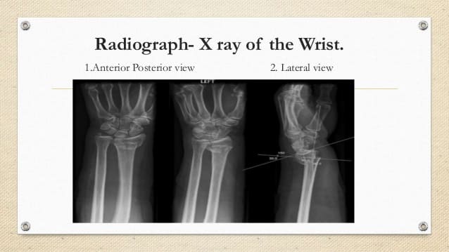
In the early phases of these injuries, radiographs are typically the main instrument utilized for assessment, diagnosis, and treatment. At the absolute least, lateral and PA views need to be taken to assess these injuries. These radiographs help to differentiate between the many types of forearm fractures and to diagnose the specific type of damage.
A Colles fracture is more likely if the patient has an extensive medical record that details the process of harm. Most of the time, a diagnosis is made exclusively by interpreting the lateral and posteroanterior images.
The classic Colles fracture has the following features:
- 2.5 centimeters, or 0.98 inches, of transverse radius fracture in front of the radiocarpal joint
- Besides the radial tilt, dorsal angulation and displacement shortening of the radius
- Loss of Ulnar inclination
- Radial angulation of the wrist
- crushing at the fracture’s location
- Over 60% of patients also had a fracture of the ulnar styloid process.
CT-scan:
The main purposes of a CT scan are to define an intraarticular fracture pattern and to facilitate surgical planning.
MRI:
` An MRI is not recommended as the initial diagnostic procedure, even if it can be utilized to assess the ligamentous or soft tissue extents of some injuries, such as TFCC, scapholunate, or lunotriquetral ligament injuries
Differential Diagnosis:
- Scapholunate ligament tear: Median nerve damage; up to 50% when ulnar styloid fx is also present; TFCC (triangular fibrocartilage complex) injuryCarpal ligament injury: most prevalent is scapholunate instability; lunotriquetral ligament
- EIP tendon transfer is typically used to treat tendon injuries and attritional EPL ruptures.
- The syndrome of compartments
- fracture of the ulnar styloid
- Instability of the Distal Radial Ulnar Joint
- Strongly correlated with distal 1/3 radial shaft fractures is the Galeazzi fracture.
Treatment of Colle’s Fracture:
The course of therapy for Colles fractures will depend on the type of fracture that is present, the patient’s age and activity level, the surgeon’s decision, and the patient’s needs regarding immobilization and return to activity. Numerous therapeutic approaches have been developed to stabilize Colles fractures and encourage bone healing as a result of their high frequency. The ultimate goal is to return the wrist to its pre-injury functioning state.
How a Colles fracture is treated depends on its severity. The conservative course of treatment for an undisplaced fracture may involve a cast alone. The cast is placed with the distal portion in palmar flexion and ulnar
Surgical procedures include internal fixation, external fixation, percutaneous pinning, and bone replacements.
Closed reduction may be necessary for a fracture that is somewhat displaced and angulated. An open reduction and internal or external fixation may be necessary for significant angulation and deformity.
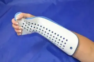
For optimal outcomes, temporarily immobilize fractures of the forearm, wrist, and hand, including Colles fractures, using a volar forearm splint.
An increase in the number of instability criteria increases the possibility of an operative treatment. After considering the patient’s age and physical needs, the fracture pattern, degree of displacement, and stability, the best course of action will be decided.
Orthopedic Treatment:
Orthopaedic Management and Care are primarily dependent on the extent of the fracture. For an undisplaced fracture, immobilization can be achieved with a cast or plaster. The cast is placed with the distal portion in palmar flexion and ulnar deviation. A moderate displaced and angulated fracture may require closed reduction.
The idea that immobilizing the wrist in dorsiflexion as opposed to palmar flexion results in less displacement and better functional status is not well-supported by the available research. For severe angulation and deformity, an open reduction and internal or external fixation can be required. For the best temporary immobilization of hand, wrist, and forearm fractures, including Colle’s fracture, use a volar forearm splint.
There are several accepted criteria for instability:
- ulnar styloid process abruption, dorsal tilt of more than 20°, comminuted fracture, intraarticular displacement of more than 1 mm, and loss of radial height of more than 2 mm
- Operative intervention is necessary if the quantity of unstable fractures is higher.
- The elderly require different treatment approaches.
- To ensure adequate healing, routine follow-up X-ray exams are necessary after one, two, and six weeks.
- Following Cast, Fixation, or Plaster Removal of Wrist Joint Stiffness, Physiotherapy Exercises are recommended.
Physical Therapy Of Colle’s Fracture:
Many patients with pain, edema, decreased strength, limited range of motion, and diminished functional abilities are seen by physiotherapists often. Following the healing of a Colles fracture, therapy is recommended to restore strength and function to the fractured wrist.
The most important thing in early therapy is wrist mobilization. This is expected to occur 7–8 weeks following the fracture. If an internal fixation device has been utilized to control the fracture, early mobilization can begin as soon as one week following surgery.
Fractures treated with external fixation should be handled carefully since the wrist is often kept in a pronated posture. The patient may be at risk for distal radioulnar joint contracture as a result of this. Other soft tissue injuries that may hinder the healing process include edema, cast impingement, infection, osteomyelitis, adherent scar, intrinsic or extrinsic muscle tightness, joint capsular tightness, neurovascular injury, ligament damage, post-traumatic arthritis
Initial Rehabilitation:
One of the primary goals of early rehabilitation is the restoration of the wrist’s natural range of motion (ROM), which can be achieved by both passive ROM and progression to active ROM. The first motions stressed are often wrist flexion and extension, working within the patient’s pain-free accessible range.
Range-of-motion exercises help to lessen the formation of adhesions and scar tissue, which often occur after surgery. It’s also essential to focus motion at the joints above and below (the elbow, fingers, and shoulder) during the entire rehabilitation process. One of the primary objectives of early rehabilitation is to decrease the degree of edema and pain in the wrist and hand region.
Extending the wrist range of motion and performing strengthening exercises are the main goals of the subsequent phase of rehabilitation for a Colles fracture. Six to eight weeks following surgery is when range of motion (ROM) for fractures treated surgically should be restored.
Some examples of ROM exercises that can be performed are as follows:
- Wrist flexion and extension
- Both ulnar and radial deviation
- Supination and Pronation
- A fist opening and closing
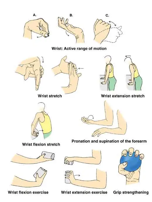
One can go toward strengthening during the sub-acute phase by performing grip squeezes with a foam ball or towel roll, or by performing all range-of-motion activities with a weight in the hand. It’s critical to strengthen the entire forearm in addition to the intrinsic and extrinsic hand muscles, progressively escalating the resistance as the person acquires strength.
At this point, progressive stretching can begin to increase the amount of accessible range of motion. Each stretch should be held for 30 to 60 seconds for three repetitions. In case the patient is unable to tolerate a slow, protracted stretch, you can utilize ten repetitions of 10 seconds each.
Acute Phase:
Paraffin wax/heat:
Heat, either a heat pack or paraffin wax, can be quite beneficial in the early stages for increasing the range of motion and reducing pain. It is often used along with cold therapy to enhance venous return.
Massage:
Two efficient massage techniques utilized in post-Colle’s fracture rehabilitation include retrograde massage to reduce swelling and massage to remove scar tissue.
One benefit is that the patient can learn how to manage independently while residing in their own home.
Cryotherapy:
Cryotherapy is a useful way to manage edema in the acute phase after trauma and throughout rehabilitation because it can help reduce blood flow by vasoconstriction, which restricts the amount of fluid leaving capillaries to the interstitial fluid. To treat edema, compression, and elevation can also be applied in addition to cryotherapy. Pain management for up to two hours following treatment can be achieved by applying cryotherapy to the affected area for ten to fifteen minutes.
Patients who are very young or very old, have an open wound, are over a superficial branch of the nerve, or have weak sensibility or mentation should not have cryotherapy.
Conditions that prohibit the use of cryotherapy include cryoglobinemia, cold urticaria, acute febrile illness, and vasospasm (such as Raynaud’s disease).
Electrical Stimulation:
Any level of rehabilitation can benefit from the adjuvant use of transcutaneous electrical nerve stimulation (TENS), a pain control therapy. On the other hand, TENS may be highly beneficial for people who are increasing wrist activity. By using a prolonged treatment session that lasts up to twenty-four hours a day, conventional (high-rate) TENS can be used to break the pain cycle.
Low-rate TENS is another electrical stimulation technique that works by stimulating the motor or nociceptive A-delta neurons to efficiently reduce pain. Low-rate TENS is an effective pain relief for up to four or five hours following therapy.
There is still a lack of conclusive research on this topic, and the results of different studies may confirm each other. However, evidence exists for the beneficial effects of electrical stimulation, especially when combined with physiotherapy exercises.
Exercise For Wrist joint:
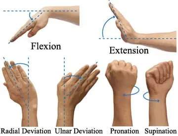
Exercise is crucial for regaining range of motion and strengthening the elbow, shoulder, wrist, and hand. The impact of wrist immobility on power and range of motion is significant. Simple hand exercises like walking up the wall can improve range of motion (ROM), while wrist exercises like writing, drawing, and tearing paper are excellent for hand strength and dexterity. To improve function and restore independence in ADL, the ability to apply opposition and pinching is essential. After a Colle’s fracture, even simple easy things like buttoning a shirt may become difficult.
Following a period of immobilization ranging from four to six weeks, your physician can decide to take off the cast and suggest physical rehabilitation. Your physical therapist may assess and measure limitations related to strength, discomfort, edema, range of motion (ROM), and other common conditions. Your physical therapist may evaluate your surgical scar tissue if you have an ORIF to reduce the fracture. He or she might also examine the functionality of your arm, wrist, and hands.
Following your initial assessment, your physical therapist is going to work with you to create a personalized therapy plan aimed at enhancing any impairments or functional restrictions you may be experiencing. Never hesitate to ask questions if you have any. Once your Colles fracture procedure, your PT can recommend a particular training regimen.
You may have experienced a significant loss of hand, wrist, and elbow movement following a Colle’s fracture. In addition, your shoulder might be tight, particularly if you have been wearing a sling. You might be required to undertake a range of motion exercises (ROM) at home for your elbow, wrist, and hand.
Strengthening Exercise:
Strength loss following a Colle’s fracture is not uncommon. Strengthening exercises for the hands, wrists, and elbows may be recommended. Once more, to receive the maximum benefit from physiotherapy, you might need to complete exercises at home.
Scar Tissue Mobilization:
If you have an ORIF operation to reduce your Colles’ fracture, the surgical incision may probably have some scarring. To assist your scar move more freely, your physical therapist could massage and mobilize the scar tissue. You can learn how to perform it on yourself from him or her as well.
Following a few weeks of physical therapy, you need to experience a decrease in pain and swelling along with an improvement in your strength and mobility. You may be finding it simpler to carry out practical tasks with your arm and hand. Although the fracture should mend completely six to eight weeks following the accident,
A Colle’s fracture, sometimes known as a broken wrist, can be frightening and painful. You could find it difficult to do simple tasks like brushing your hair, and clothing, or feeding yourself with your hands and arms. It might be impossible for you to carry out your work or enjoy leisure activities. To ensure that you can rapidly and safely return to your regular activities, your physical therapist can assist you in increasing your functional mobility.
Complication:
Usually, wrist stiffness is the most common adverse effect. It should get better after you take off your cast after a month or two. Other issues to consider include:-
Excessive pressure inside and around your muscles can cause compartment syndrome, a painful condition.
A malunion is when the bones are unable to come together.
Your hand muscles become paralyzed when you have median nerve palsy, making it impossible for you to touch your fingers or flex your thumb.
When you have carpal tunnel syndrome, your wrist nerves are compressed. The symptoms include weakness, numbness, and pain in the hands and wrists.
Hand, foot, arm, or leg burning pain is a sign of reflex sympathetic dystrophy.
Your wrist could develop secondary osteoarthritis. Among the symptoms include pain and swelling in the deformed joint.
Prognosis:
The degree of the damage and the severity of any aftereffects determine the prognosis for a Colles fracture. Early and appropriate reduction, casting, splinting, and follow-up care from an orthopedist can prevent complications. Orthopedic consultation is necessary immediately for significant injuries such as open fractures, injuries involving neurovascular impairment, or symptoms of compartment syndrome.
Serious damage often requires surgery to treat. The age of the patient also affects the prognosis; younger patients are more likely to benefit from bone remodeling, whereas elderly patients are less likely to do so.
Conclusion
A Colles fracture is an extremely painful and dangerous injury. For treatment, you should visit the emergency room if you have a painful wrist injury. To help you recover, your healthcare professionals will evaluate your injuries, realign your bones, and put you in a cast or splint.
Consult your medical professionals regarding osteoporosis. It may indicate that you have a fractured wrist. Your doctors will address any concerns you have and go over additional diagnostic procedures and if required, treatment options.
FAQs
What joint is a fracture of the Colles?
One of the most common wrist injuries is Colles fractures, which are caused by a combination of distal radius fractures and dorsal displacement.
With a Colles fracture, which structures are harmed?
Colles fractures are the most common cause of wrist injuries. It usually occurs when a hand that is extended falls. The impact of the fall fractures the distal part of the radius, causing it to shift into the typical “silver fork” posture.
In cases of Colles fracture, what kind of splint is used?
For the best temporary immobilization of hand, wrist, and forearm fractures, including Colles fractures, use a volar forearm splint.
What is the Colle’s fracture known by another name?
Colles’ fractures are the most prevalent type of wrist fracture. A Colles’ fracture affects the larger of the two wrist bones, the radius, and is also referred to as a “distal radius fracture.”
What is the location of a Colles fracture?
A Colles fracture is a break in the wrist’s radius. The name of the surgeon who first described it was given to it. Usually, the break is visible around an inch (2.5 cm) below the wrist’s connecting point.
References
- Solanki, G. (2024, January 4). Colles Fracture – Symptoms, Treatment, and Recovery Time. Mobile Physiotherapy Clinic. https://mobilephysiotherapyclinic.in/colles-fracture
- Summers, K., Mabrouk, A., & Fowles, S. M. (2023, July 31). Colles Fracture. StatPearls – NCBI Bookshelf. https://www.ncbi.nlm.nih.gov/books/NBK553071/
- Dhameliya, N., & Dhameliya, N. (2023, April 27). Colle’s fracture: – Causes, Symptoms, Treatment – Samarpan. Samarpan Physiotherapy Clinic. https://samarpanphysioclinic.com/colles-fracture/




2 Comments