Medial Collateral Ligament (MCL)
Introduction
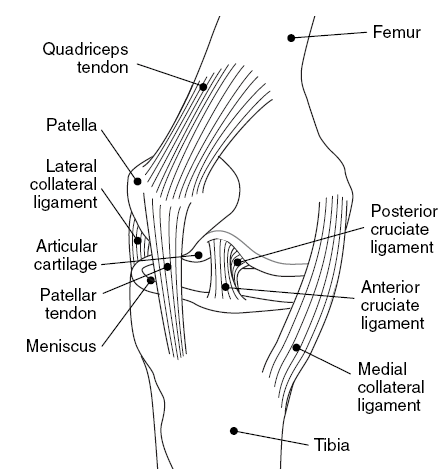
The Medial Collateral Ligament (MCL) is a band of tissue located on the inner side of the knee, connecting the femur (thigh bone) to the tibia (shin bone). It provides stability by preventing excessive inward movement of the knee joint.
MCL injuries can range from minor sprains to full tears and can be caused by direct trauma or abrupt twisting motions. Although surgery may be necessary in more severe cases, MCL injuries are usually treated with rest, ice, compression, and physical therapy.
Anatomy of the medial collateral ligament
Slightly posteriorly on the medial side of the knee joint, it is a wide, flat, membranous band. Proximally, it is connected to the femur’s medial epicondyle just behind the adductor tubercle; below, it is connected to the tibia’s medial condyle and the medial surface of its body. Pressures that might occur in valgus deformity are prevented by pushing the knee medially.
The posterior portion of the ligament is placed into the tibia above the semimembranosus muscle groove; its fibers are short and downward inclining.
About 10 centimeters long, the anterior portion of the ligament is a flattened band that slopes forward as it descends.
Approximately 2.5 centimeters below the condyle’s level, it is placed into the medial surface of the tibia.
The sartorius, gracilis, and semitendinosus muscles’ linked tendons, known as the pes anserine, cross over the bottom portion of the MCL; a bursa sits in between.
Structure of the medial collateral ligament
A robust, fibrous band of tissue that runs down the interior of the knee joint is called the MCL. It gives the ligament strength and flexibility and is made up of many layers of crisscrossed collagen fibers.
The medial epicondyle of the femur, a bony protuberance on the inside of the thigh bone, is where the MCL connects at its upper end. After there, it descends and joins the tibia right below the joint line. Additionally, the lower end of the MCL connects to the medial malleolus, a little ankle bone.
The superficial and deep layers are the two primary sections that make up the MCL. Resisting forces that push the knee inward (valgus stress) is the responsibility of the superficial layer, which is found on the outside of the ligament. The deep layer, on the other hand, is found on the ligament’s inner surface and is responsible for resisting forces that cause the knee to twist or rotate.
Furthermore, in addition to its primary role of stabilizing the knee joint, the MCL also helps to maintain the patella’s (kneecap) proper alignment and tracking. During weight-bearing exercises, it also aids in the distribution of stresses and weight over the knee joint.
MCL injuries can significantly affect a person’s capacity to perform daily tasks, participate in sports, and engage in other physical activities. In general, the MCL’s structure is essential for preserving the stability and function of the knee joint.
Purpose of the medial collateral ligament
The MCL’s primary function is to stabilize the knee joint, especially during side-to-side motions.
The MCL serves as the knee’s main barrier against forces that drive the knee outward, as those that could happen during a football tackle or a skiing collision. It also aids in preventing the knee from moving too much inward, which could harm the knee’s joint surfaces and other internal components.
In addition, the MCL contributes to shock absorption by distributing forces across the knee joint during weight-bearing exercises including running, leaping, and walking.
The MCL is susceptible to injury from overuse or abrupt impact, just like other knee ligaments. An MCL injury can cause discomfort, stiffness, edema, and trouble bending or straightening the knee. The knee may feel unsteady or give way when moving in more severe situations.
When it comes to maintaining stability and function during a range of activities, the MCL is a crucial component of the knee joint. Maintaining one’s mobility and quality of life can be facilitated by knowing its function and how to avoid and treat injuries.
Blood supply of the medial collateral ligament
The MCL’s blood flow is critical to its post-injury healing and recuperation. The medial superior genicular artery, medial inferior genicular artery, and descending genicular artery are among the several blood vessels that supply the MCL.
The popliteal artery is the source of the medial superior genicular artery, which supplies blood to the top part of the MCL. Blood is supplied to the lower part of the MCL via the medial inferior genicular artery, which likewise emerges from the popliteal artery. The MCL receives additional blood flow from the descending genicular artery, which emerges from the femoral artery.
The MCL also gets blood flow from tiny vessels that pierce its surface in addition to these arteries. Following an injury, these vessels aid in the ligament’s nourishment and healing.
The ligament’s blood flow may be interrupted by an MCL injury, which could delay the healing process. Thus, it’s critical to take action to increase blood flow to the wounded area, such as by engaging in physical therapy exercises and other rehabilitation techniques.
Ultimately, it is critical to understand the MCL’s blood supply and how to manage injuries and encourage the healing of this crucial knee joint structure.
Symptoms of the medial collateral ligament
When the ligament is overstretched and the valgus force is too high for it to withstand, the MCL is injured. A sudden change in direction, twisting the knee while the foot is fixed, landing incorrectly from a jump, or—most frequently—a blunt force injury to the knee, like in a football tackle, can all cause this. Usually, the incident must occur quickly. You are more likely to sustain a ligament sprain or tear if you have weak muscles or poor coordination.
SIGN & SYMPTOMS
- The inside of the knee is sensitive.
- The knee is bruised.
- A popping sound when hurt
- The inner area of your knee hurts and feels painful.
- Knee joint swelling
- A sensation that when you put weight on your knee, it will fail
- knee joint locking or catching
- Knee joint Stiffness
CLASSIFICATION
- Grade 1: Mild pain and some tenderness.
- Grade 2: Significant pain and tenderness at the inside of the knee; noticeable looseness when manipulated by hand; and, in certain situations, edema.
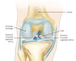
- Grade 3: Significant knee pain and tenderness, along with some swelling and noticeable joint instability. When the doctor swings your leg around, the knee opens up about 1 centimeter, which is somewhat less than half an inch. Anterior cruciate ligament tears frequently coexist with grade 3 MCL tears.
Injuries of the medial collateral ligament
There are several ways to injure the MCL, including:
- Direct impact: The MCL may stretch or tear if the outside of the knee is struck directly.
- Twisting motion: The knee may rotate inward due to abrupt twisting or pivoting motions, which could strain the MCL and result in a tear.
- Hyperextension: The MCL may be strained or torn when the knee is pressed past its typical range of motion, as happens after a fall or landing from a jump.
- Overuse: The MCL may become inflamed and eventually injured as a result of repetitive stress from activities like running or leaping.
- Poor landing technique: When landing from a jump or changing direction, poor technique can increase the risk of injury by putting too much strain on the MCL.
- Sports-related injuries: Because of the possibility of collisions and abrupt direction changes, contact sports like basketball, football, and soccer are more likely to cause MCL injuries.
- Car crashes: Car crashes or other stressful incidents that result in a quick impact on the knee can occasionally cause MCL tear.
Treatment of the medial collateral ligament
Conservative treatment
Non-surgical techniques are used in the conservative treatment of medial collateral ligament (MCL) injuries to encourage healing and lessen discomfort and inflammation. The steps involved in treating MCL conservatively are as follows:
- Rest: To aid in the healing process, the injured knee must be rested. Any activity that makes the patient’s knee hurt or uncomfortable should be avoided.
- Ice: You can lessen discomfort and inflammation in your knee by using ice for 20 to 30 minutes, three to four times a day.
- Compression: Supporting the knee and lowering swelling can be achieved by using a knee brace or compression bandage.
- Elevation: Raising the affected knee above the level of the heart can aid in promoting blood flow to the area and reducing edema.
- Medication: Ibuprofen and acetaminophen are examples of over-the-counter pain relievers that can be very helpful in reducing pain and inflammation.
- Physical treatment: Exercises used in physical therapy can assist in strengthening the knee’s surrounding muscles and encourage MCL recovery.
- Activity modification: Reducing knee-stressing activities like jogging and leaping can help stop new injuries and speed up recovery.
- Gradual return to activity: Resuming regular activities after the knee has healed can assist avoid re-injury.
Rest, ice, compression, elevation, medication, physical therapy, activity adjustment, and a gradual return to exercise are all part of the conservative treatment of MCL. For the best healing and recovery, it’s critical to adhere to the treatment plan that a doctor prescribes.
Physiotherapy treatment
The goal of physiotherapy for MCL (Medial Collateral Ligament) injuries is to increase the range of motion, decrease pain, and encourage recovery through a variety of exercises and procedures. The steps in physiotherapy treatment for MCL are as follows:
- Evaluation: The physiotherapist will determine the best direction of action after determining the severity of the MCL injury.
- To lessen pain and swelling, the physiotherapist may provide ice and compression to the knee before beginning the exercises.
- Exercises for range of motion: To increase the patient’s knee’s range of motion, the physiotherapist will lead them through some exercises. These could consist of passive range-of-motion exercises, active range-of-motion exercises, and mild stretches.
- Strengthening exercises: The physiotherapist will lead the patient through a series of exercises to strengthen the muscles surrounding the knee when the range of motion has improved. Examples of these could be exercises that strengthen the hamstrings, calf muscles, and quadriceps.
- Exercises for balance and stability: To help the patient better support their weight on the injured knee, the physiotherapist may also incorporate these exercises.
- Exercises that enhance a patient’s awareness of the location and motion of their knee joint are known as proprioceptive training. This may lessen the chance of getting hurt again.
- Gait training: To ensure appropriate alignment and lessen strain on the knee joint, the physiotherapist may also work with the patient on their gait.
- Gradual return to activity: The physiotherapist will help the patient gradually resume their regular activities after the knee has recovered. To make sure the patient is prepared to safely resume their sport, this may involve sports-specific training.
A variety of range-of-motion exercises, strengthening exercises, balance and stability exercises, proprioceptive training, gait training, and a gradual return to activity are all part of the physiotherapy treatment for MCL. For the best healing and rehabilitation, it’s critical to adhere to the treatment plan that a physiotherapist prescribes.
Risk factors of the medial collateral ligament
MCL injuries may result from several causes, such as:
- Sports: MCL injuries are most common in sports requiring quick direction changes, including basketball, football, soccer, and skiing.
- Direct impact: An MCL tear may result from a direct hit to the outside of the knee.
- Overuse: An MCL injury can result from repetitive strain on the MCL from exercises like running or leaping.
- Age: Our body’s ligaments become less flexible and more vulnerable to damage as we age.
- Obesity: Being overweight increases the risk of MCL injury by placing more strain on the knee joint.
- Prior knee injuries: An MCL injury may be more likely to occur if you have had a prior knee injury, such as an ACL rupture.
- Poor conditioning: An increased risk of MCL injury might result from weak muscles surrounding the knee joint.
- Poor technique: During exercise or sports, using poor technique can raise your risk of suffering an MCL injury.
To avoid MCL injuries, it is critical to understand these risk factors and implement the necessary safety measures. Maintaining a healthy weight, warming up and stretching properly before activity, utilizing appropriate techniques during sports, and working with a physiotherapist to increase knee joint muscle strength and flexibility are a few examples of this.
How to reduce the chance of medial collateral ligament injury
Some safety measures are necessary to lower the chance of suffering an MCL injury. MCL injuries can be avoided in the following ways:
- Warming up and stretching: It’s crucial to properly warm up and stretch before engaging in any sports or workout. This lowers the chance of injury by preparing the muscles, tendons, and ligaments for physical exercise.
- Use the right technique: Sports requiring quick direction changes, such as basketball, football, soccer, and skiing, are particularly prone to MCL injuries. For instance, it’s crucial to pivot on the balls of your feet rather than twisting your knee when playing sports like basketball or football that need quick direction changes.
- Wear the right gear: You can lower your risk of MCL injuries by wearing the right gear, such as braces, knee pads, or supportive shoes.
- Keep your weight in check: Being overweight increases the risk of MCL injury by placing additional strain on the knee joint. Reducing this risk can be greatly aided by maintaining a healthy weight.
- Develop stronger knee muscles: MCL injuries can be more likely to occur in people with weak knee muscles. One way to lower this risk is to work with a physiotherapist to increase knee joint muscle strength and flexibility.
- Avoid overuse: MCL injuries can result from repetitive strain on the MCL from exercises like jogging or leaping. Avoiding misuse and allowing your body to rest and recuperate is crucial.
- Be cautious when recovering from an injury: It’s crucial to use caution when returning to sports or physical activity if you have previously suffered a knee injury, such as an ACL tear. A safe and progressive return to exercise plan created in collaboration with a physiotherapist can help lower the chance of further injury.
In summary, preventing MCL injuries entails taking the right precautions, including warming up, using the right technique, wearing the right equipment, keeping a healthy weight, strengthening the muscles surrounding the knee joint, avoiding overuse, and exercising caution when recovering from an injury. These recommendations will help you maintain strong, healthy knees and lower your chance of MCL injuries.
Summary
The ligament on the inside of the knee is called the medial collateral ligament, or MCL. It helps to minimize the knee’s excessive inward movement and provides stability to the knee joint. Overuse injuries, direct trauma to the knee, and abrupt twisting or bending of the knee can all result in MCL injuries.
Pain, swelling, and trouble moving the knee are all signs of an MCL injury. Physical therapy, elevation, compression, ice, rest, and in certain situations, surgery, can all be used to treat MCL injuries. The degree of the injury and the treatment strategy can affect how long it takes to recover from an MCL injury.
FAQs
The MCL: What is it?
On the inside of the knee, the medial collateral ligament (MCL) serves as a ligament that joins the thigh and shin bones.
What causes MCL injuries?
MCL injuries can happen when the knee suddenly twists or is struck, as happens in sports or other strenuous activities.
What features distinguish an MCL injury?
Pain, swelling, stiffness, and trouble bearing weight on the injured leg are all signs of an MCL injury.
What is the treatment for MCL injuries?
Rest, ice, compression, and elevation (RICE), physical therapy, and/or bracing are often used treatments for MCL injuries. In extreme circumstances, surgery can be required.
What is the average recovery period following an MCL injury?
Depending on the injury’s impact, recovery times for MCL injuries can vary. While more severe injuries may take several months to fully recover, milder injuries may heal in a few weeks.
References
- Patel, D. (2023, December 13). Medial collateral ligament – structure, function, injury. Mobile Physiotherapy Clinic. https://mobilephysiotherapyclinic.in/medial-collateral-ligament/
- Dhameliya, N. (2024, December 25). MEDIAL COLLATERAL LIGAMENT – anatomy, function. Samarpan Physiotherapy Clinic. https://samarpanphysioclinic.com/medial-collateral-ligament/
One Comment